1KME

CRYSTAL STRUCTURE OF BACTERIORHODOPSIN CRYSTALLIZED FROM BICELLES


Experimental Data Snapshot

  • Method: X-RAY DIFFRACTION
  • Resolution: 2.00 Å
  • R-Value Free: 0.275 
  • R-Value Work: 0.263 
  • R-Value Observed: 0.263 

wwPDB Validation   3D Report Full Report


This is version 1.4 of the entry. See complete history


Literature

Bicelle crystallization: a new method for crystallizing membrane proteins yields a monomeric bacteriorhodopsin structure.

Faham, S.Bowie, J.U.

(2002) J Mol Biol 316: 1-6

  • DOI: https://doi.org/10.1006/jmbi.2001.5295
  • Primary Citation of Related Structures:  
    1KME

  • PubMed Abstract: 

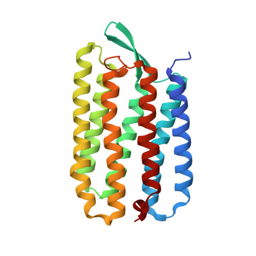
    Obtaining crystals of membrane proteins that diffract to high resolution remains a major stumbling block in structure determination. Here we present a new method for crystallizing membrane proteins from a bicelle forming lipid/detergent mixture. The method is flexible and simple to use. As a test case, bacteriorhodopsin (bR) from Halobacterium salinarum was crystallized from a bicellar solution, yielding a new bR crystal form. The crystals belong to space group P2(1) with unit cell dimensions of a=45.0 A, b=108.9 A, c=55.9 A, beta=113.58 degrees and a dimeric asymmetric unit. The structure was solved by molecular replacement and refined at 2.0 A resolution. In all previous bR structures the protein is organized as a parallel trimer, but in the crystals grown from bicelles, the individual bR subunits are arranged in an antiparallel fashion.


  • Organizational Affiliation

    Department of Chemistry and Biochemistry, University of California, Los Angeles, CA 90095-1570, USA.


Macromolecules
Find similar proteins by:  (by identity cutoff)  |  3D Structure
Entity ID: 1
MoleculeChains Sequence LengthOrganismDetailsImage
Bacteriorhodopsin
A, B
231Halobacterium salinarumMutation(s): 0 
Membrane Entity: Yes 
UniProt
Find proteins for P02945 (Halobacterium salinarum (strain ATCC 700922 / JCM 11081 / NRC-1))
Explore P02945 
Go to UniProtKB:  P02945
Entity Groups  
Sequence Clusters30% Identity50% Identity70% Identity90% Identity95% Identity100% Identity
UniProt GroupP02945
Sequence Annotations
Expand
  • Reference Sequence
Experimental Data & Validation

Experimental Data

  • Method: X-RAY DIFFRACTION
  • Resolution: 2.00 Å
  • R-Value Free: 0.275 
  • R-Value Work: 0.263 
  • R-Value Observed: 0.263 
  • Space Group: P 1 21 1
Unit Cell:
Length ( Å )Angle ( ˚ )
a = 45α = 90
b = 108.9β = 113.58
c = 55.9γ = 90
Software Package:
Software NamePurpose
AMoREphasing
CNSrefinement
DENZOdata reduction
SCALEPACKdata scaling

Structure Validation

View Full Validation Report


Entry History 

Deposition Data

Revision History  (Full details and data files)

  • Version 1.0: 2002-02-13
    Type: Initial release
  • Version 1.1: 2008-04-27
    Changes: Version format compliance
  • Version 1.2: 2011-07-13
    Changes: Version format compliance
  • Version 1.3: 2020-07-29
    Type: Remediation
    Reason: Carbohydrate remediation
    Changes: Data collection, Derived calculations, Structure summary
  • Version 1.4: 2023-08-16
    Changes: Data collection, Database references, Refinement description, Structure summary