Side-chain repacking calculations for predicting structures and stabilities of heterodimeric coiled coils.
Keating, A.E., Malashkevich, V.N., Tidor, B., Kim, P.S.(2001) Proc Natl Acad Sci U S A 98: 14825-14830
- PubMed: 11752430
- DOI: https://doi.org/10.1073/pnas.261563398
- Primary Citation of Related Structures:
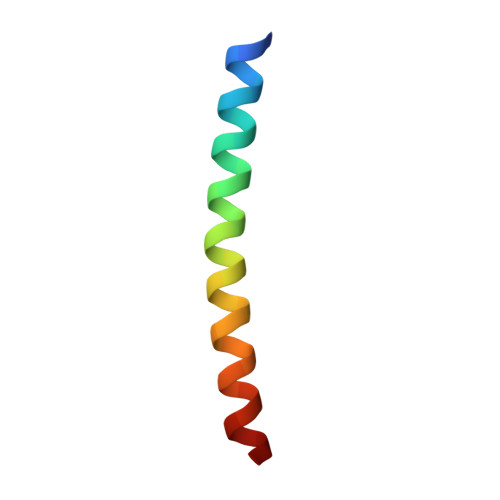
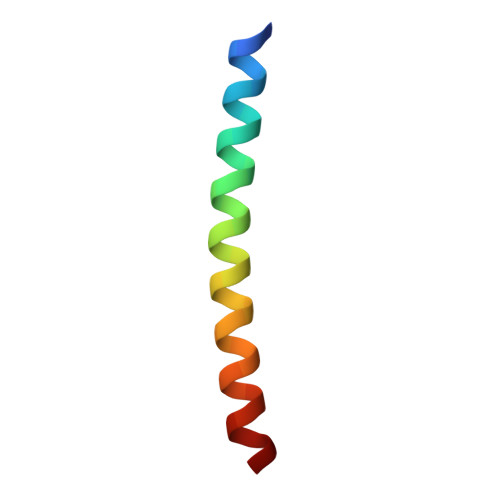
1KD8, 1KD9, 1KDD - PubMed Abstract:
An important goal in biology is to predict from sequence data the high-resolution structures of proteins and the interactions that occur between them. In this paper, we describe a computational approach that can make these types of predictions for a series of coiled-coil dimers. Our method comprises a dual strategy that augments extensive conformational sampling with molecular mechanics minimization. To test the performance of the method, we designed six heterodimeric coiled coils with a range of stabilities and solved x-ray crystal structures for three of them. The stabilities and structures predicted by the calculations agree very well with experimental data: the average error in unfolding free energies is <1 kcal/mol, and nonhydrogen atoms in the predicted structures superimpose onto the experimental structures with rms deviations <0.7 A. We have also tested the method on a series of homodimers derived from vitellogenin-binding protein. The predicted relative stabilities of the homodimers show excellent agreement with previously published experimental measurements. A critical step in our procedure is to use energy minimization to relax side-chain geometries initially selected from a rotamer library. Our results show that computational methods can predict interaction specificities that are in good agreement with experimental data.
Organizational Affiliation:
Whitehead Institute for Biomedical Research, Howard Hughes Medical Institute, Department of Biology, Massachusetts Institute of Technology, Nine Cambridge Center, Cambridge, MA 02142, USA. keating@mit.edu