1JZD

DsbC-DsbDalpha complex


Experimental Data Snapshot

  • Method: X-RAY DIFFRACTION
  • Resolution: 2.30 Å
  • R-Value Free: 0.293 
  • R-Value Work: 0.234 

wwPDB Validation   3D Report Full Report


This is version 1.4 of the entry. See complete history


Literature

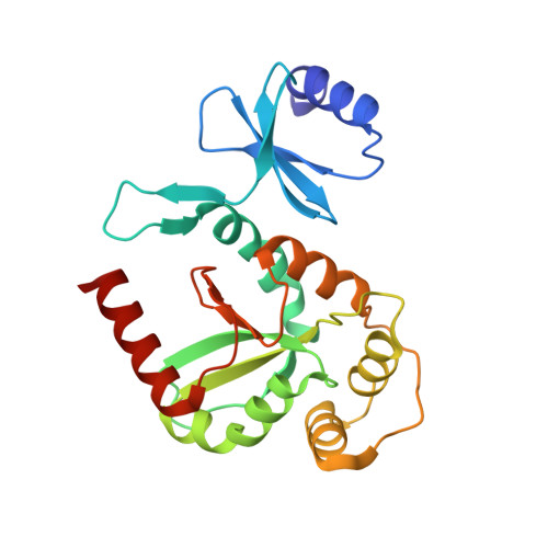
The Disulfide Bond Isomerase DsbC is Activated by an Immunoglobulin-fold Thiol Oxidoreductase: Crystal structure of the DsbC-DsbDalpha complex.

Haebel, P.W.Goldstone, D.Katzen, F.Beckwith, J.Metcalf, P.

(2002) EMBO J 21: 4774-4784

  • DOI: https://doi.org/10.1093/emboj/cdf489
  • Primary Citation of Related Structures:  
    1JPE, 1JZD, 1JZO

  • PubMed Abstract: 

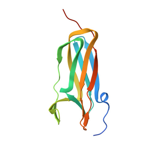
    The Escherichia coli disulfide bond isomerase DsbC rearranges incorrect disulfide bonds during oxidative protein folding. It is specifically activated by the periplasmic N-terminal domain (DsbDalpha) of the transmembrane electron transporter DsbD. An intermediate of the electron transport reaction was trapped, yielding a covalent DsbC-DsbDalpha complex. The 2.3 A crystal structure of the complex shows for the first time the specific interactions between two thiol oxidoreductases. DsbDalpha is a novel thiol oxidoreductase with the active site cysteines embedded in an immunoglobulin fold. It binds into the central cleft of the V-shaped DsbC dimer, which assumes a closed conformation on complex formation. Comparison of the complex with oxidized DsbDalpha reveals major conformational changes in a cap structure that regulates the accessibility of the DsbDalpha active site. Our results explain how DsbC is selectively activated by DsbD using electrons derived from the cytoplasm.


  • Organizational Affiliation

    School of Biological Sciences, University of Auckland, Private Bag 92019, Auckland, New Zealandand.


Macromolecules
Find similar proteins by:  (by identity cutoff)  |  3D Structure
Entity ID: 1
MoleculeChains Sequence LengthOrganismDetailsImage
thiol:disulfide interchange protein dsbc
A, B
220Escherichia coliMutation(s): 1 
Gene Names: dsbC
UniProt
Find proteins for P0AEG6 (Escherichia coli (strain K12))
Explore P0AEG6 
Go to UniProtKB:  P0AEG6
Entity Groups  
Sequence Clusters30% Identity50% Identity70% Identity90% Identity95% Identity100% Identity
UniProt GroupP0AEG6
Sequence Annotations
Expand
  • Reference Sequence
Find similar proteins by:  (by identity cutoff)  |  3D Structure
Entity ID: 2
MoleculeChains Sequence LengthOrganismDetailsImage
thiol:disulfide interchange protein dsbd132Escherichia coliMutation(s): 1 
Gene Names: dsbD
UniProt
Find proteins for P36655 (Escherichia coli (strain K12))
Explore P36655 
Go to UniProtKB:  P36655
Entity Groups  
Sequence Clusters30% Identity50% Identity70% Identity90% Identity95% Identity100% Identity
UniProt GroupP36655
Sequence Annotations
Expand
  • Reference Sequence
Experimental Data & Validation

Experimental Data

  • Method: X-RAY DIFFRACTION
  • Resolution: 2.30 Å
  • R-Value Free: 0.293 
  • R-Value Work: 0.234 
  • Space Group: P 41 21 2
Unit Cell:
Length ( Å )Angle ( ˚ )
a = 68.91α = 90
b = 68.91β = 90
c = 230.336γ = 90
Software Package:
Software NamePurpose
MOSFLMdata reduction
SCALAdata scaling
MOLREPphasing
CNSrefinement
CCP4data scaling

Structure Validation

View Full Validation Report


Entry History 

Deposition Data

Revision History  (Full details and data files)

  • Version 1.0: 2003-03-08
    Type: Initial release
  • Version 1.1: 2008-04-27
    Changes: Version format compliance
  • Version 1.2: 2011-07-13
    Changes: Version format compliance
  • Version 1.3: 2021-10-27
    Changes: Data collection, Database references
  • Version 1.4: 2023-08-16
    Changes: Data collection, Refinement description