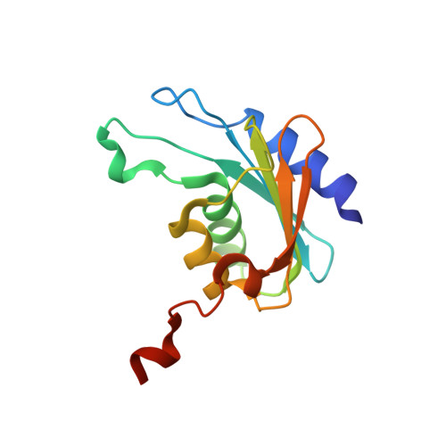
Crystal structure of the tetrameric cytidine deaminase from Bacillus subtilis at 2.0 A resolution.
Johansson, E., Mejlhede, N., Neuhard, J., Larsen, S.(2002) Biochemistry 41: 2563-2570
- PubMed: 11851403
- DOI: https://doi.org/10.1021/bi011849a
- Primary Citation of Related Structures:
1JTK - PubMed Abstract:
Cytidine deaminases (CDA, EC 3.5.4.5) are zinc-containing enzymes in the pyrimidine salvage pathway that catalyze the formation of uridine and deoxyuridine from cytidine and deoxycytidine, respectively. Two different classes have been identified in the CDA family, a homodimeric form (D-CDA) with two zinc ions per dimer and a homotetrameric form (T-CDA) with four zinc ions per tetramer. We have determined the first structure of a T-CDA from Bacillus subtilis. The active form of T-CDA is assembled of four identical subunits with one active site apiece. The subunit of D-CDA is composed of two domains each exhibiting the same fold as the T-CDA subunits, but only one of them contains zinc in the active site. The similarity results in a conserved structural core in the two CDA forms. An intriguing difference between the two CDA structures is the zinc coordinating residues found at the N-terminal of two alpha-helices: three cysteine residues in the tetrameric form and two cysteine residues and one histidine residue in the dimeric form. The role of the zinc ion is to activate a water molecule and thereby generate a hydroxide ion. How the zinc ion in T-CDA surrounded with three negatively charged residues can create a similar activity of T-CDA compared to D-CDA has been an enigma. However, the structure of T-CDA reveals that the negative charge caused by the three ligands is partly neutralized by (1) an arginine residue hydrogen-bonded to two of the cysteine residues and (2) the dipoles of two alpha-helices.
Organizational Affiliation:
Centre for Crystallographic Studies, Department of Chemistry, University of Copenhagen, Universitetsparken 5, DK-2100 Copenhagen Ø, Denmark.