1JOP

YHCH protein (HI0227)


Experimental Data Snapshot

  • Method: X-RAY DIFFRACTION
  • Resolution: 2.60 Å
  • R-Value Work: 0.185 
  • R-Value Observed: 0.185 

wwPDB Validation   3D Report Full Report


This is version 1.3 of the entry. See complete history


Literature

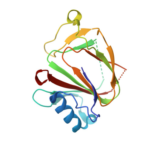
Crystal structure of the bacterial YhcH protein indicates a role in sialic acid catabolism.

Teplyakov, A.Obmolova, G.Toedt, J.Galperin, M.Y.Gilliland, G.L.

(2005) J Bacteriol 187: 5520-5527

  • DOI: https://doi.org/10.1128/JB.187.16.5520-5527.2005
  • Primary Citation of Related Structures:  
    1JOP, 1S4C

  • PubMed Abstract: 

    The yhcH gene is part of the nan operon in bacteria that encodes proteins involved in sialic acid catabolism. Determination of the crystal structure of YhcH from Haemophilus influenzae was undertaken as part of a structural genomics effort in order to assist with the functional assignment of the protein. The structure was determined at 2.2-A resolution by multiple-wavelength anomalous diffraction. The protein fold is a variation of the double-stranded beta-helix. Two antiparallel beta-sheets form a funnel opened at one side, where a putative active site contains a copper ion coordinated to the side chains of two histidine and two carboxylic acid residues. A comparison to other proteins with a similar fold and analysis of the genomic context suggested that YhcH may be a sugar isomerase involved in processing of exogenous sialic acid.


  • Organizational Affiliation

    Center for Advanced Research in Biotechnology, University of Maryland Biotechnology Institute and National Institute of Standards and Technology, Rockville, Maryland, USA. ATeplyak@cntus.jnj.com


Macromolecules
Find similar proteins by:  (by identity cutoff)  |  3D Structure
Entity ID: 1
MoleculeChains Sequence LengthOrganismDetailsImage
Yhch protein
A, B, C, D
155Haemophilus influenzaeMutation(s): 0 
UniProt
Find proteins for P44583 (Haemophilus influenzae (strain ATCC 51907 / DSM 11121 / KW20 / Rd))
Explore P44583 
Go to UniProtKB:  P44583
Entity Groups  
Sequence Clusters30% Identity50% Identity70% Identity90% Identity95% Identity100% Identity
UniProt GroupP44583
Sequence Annotations
Expand
  • Reference Sequence
Experimental Data & Validation

Experimental Data

  • Method: X-RAY DIFFRACTION
  • Resolution: 2.60 Å
  • R-Value Work: 0.185 
  • R-Value Observed: 0.185 
  • Space Group: P 1 21 1
Unit Cell:
Length ( Å )Angle ( ˚ )
a = 43.1α = 90
b = 152.3β = 113.6
c = 53.4γ = 90
Software Package:
Software NamePurpose
DENZOdata reduction
SCALEPACKdata scaling
MLPHAREphasing
REFMACrefinement

Structure Validation

View Full Validation Report


Entry History 

Deposition Data

Revision History  (Full details and data files)

  • Version 1.0: 2003-06-17
    Type: Initial release
  • Version 1.1: 2008-04-27
    Changes: Version format compliance
  • Version 1.2: 2011-07-13
    Changes: Version format compliance
  • Version 1.3: 2024-02-07
    Changes: Data collection, Database references, Derived calculations