1JKE

D-Tyr tRNATyr deacylase from Escherichia coli


Experimental Data Snapshot

  • Method: X-RAY DIFFRACTION
  • Resolution: 1.55 Å
  • R-Value Free: 0.228 
  • R-Value Work: 0.209 
  • R-Value Observed: 0.209 

wwPDB Validation   3D Report Full Report


This is version 1.4 of the entry. See complete history


Literature

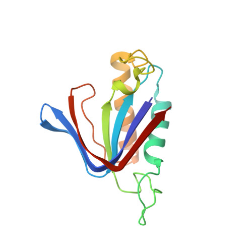
Structure of crystalline D-Tyr-tRNA(Tyr) deacylase. A representative of a new class of tRNA-dependent hydrolases.

Ferri-Fioni, M.L.Schmitt, E.Soutourina, J.Plateau, P.Mechulam, Y.Blanquet, S.

(2001) J Biol Chem 276: 47285-47290

  • DOI: https://doi.org/10.1074/jbc.M106550200
  • Primary Citation of Related Structures:  
    1JKE

  • PubMed Abstract: 

    Cell growth inhibition by several d-amino acids can be explained by an in vivo production of d-aminoacyl-tRNA molecules. Escherichia coli and yeast cells express an enzyme, d-Tyr-tRNA(Tyr) deacylase, capable of recycling such d-aminoacyl-tRNA molecules into free tRNA and d-amino acid. Accordingly, upon inactivation of the genes of the above deacylases, the toxicity of d-amino acids increases. Orthologs of the deacylase are found in many cells. In this study, the crystallographic structure of dimeric E. coli d-Tyr-tRNA(Tyr) deacylase at 1.55 A resolution is reported. The structure corresponds to a beta-barrel closed on one side by a beta-sheet lid. This barrel results from the assembly of the two subunits. Analysis of the structure in relation with sequence homologies in the orthologous family suggests the location of the active sites at the carboxy end of the beta-strands. The solved structure markedly differs from those of all other documented tRNA-dependent hydrolases.


  • Organizational Affiliation

    Laboratoire de Biochimie, Unité Mixte de Recherche 7654, CNRS-Ecole Polytechnique, F-91128 Palaiseau cedex, France.


Macromolecules
Find similar proteins by:  (by identity cutoff)  |  3D Structure
Entity ID: 1
MoleculeChains Sequence LengthOrganismDetailsImage
D-Tyr-tRNATyr deacylase
A, B, C, D
145Escherichia coliMutation(s): 0 
Gene Names: YIHZ or B3887
UniProt
Find proteins for P0A6M4 (Escherichia coli (strain K12))
Explore P0A6M4 
Go to UniProtKB:  P0A6M4
Entity Groups  
Sequence Clusters30% Identity50% Identity70% Identity90% Identity95% Identity100% Identity
UniProt GroupP0A6M4
Sequence Annotations
Expand
  • Reference Sequence
Experimental Data & Validation

Experimental Data

  • Method: X-RAY DIFFRACTION
  • Resolution: 1.55 Å
  • R-Value Free: 0.228 
  • R-Value Work: 0.209 
  • R-Value Observed: 0.209 
  • Space Group: C 1 2 1
Unit Cell:
Length ( Å )Angle ( ˚ )
a = 118.3α = 90
b = 82.93β = 128.29
c = 83.51γ = 90
Software Package:
Software NamePurpose
MOSFLMdata reduction
SCALAdata scaling
SHARPphasing
CNSrefinement
CCP4data scaling

Structure Validation

View Full Validation Report


Entry History 

Deposition Data

Revision History  (Full details and data files)

  • Version 1.0: 2002-01-25
    Type: Initial release
  • Version 1.1: 2008-04-27
    Changes: Version format compliance
  • Version 1.2: 2011-07-13
    Changes: Version format compliance
  • Version 1.3: 2011-11-16
    Changes: Atomic model
  • Version 1.4: 2024-02-07
    Changes: Data collection, Database references, Derived calculations