Structural analyses of nucleotide binding to an aminoglycoside phosphotransferase.
Burk, D.L., Hon, W.C., Leung, A.K., Berghuis, A.M.(2001) Biochemistry 40: 8756-8764
- PubMed: 11467935
- DOI: https://doi.org/10.1021/bi010504p
- Primary Citation of Related Structures:
1J7I, 1J7L, 1J7U - PubMed Abstract:
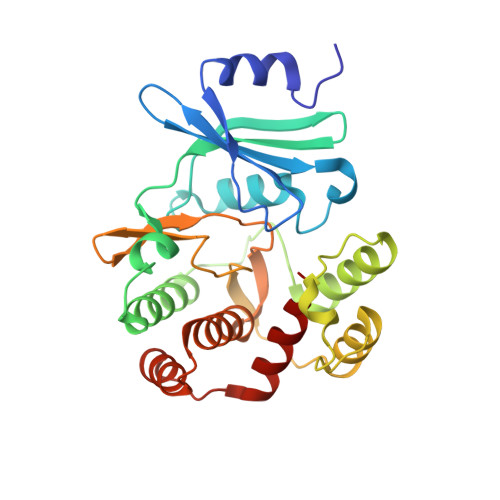
3',5"-Aminoglycoside phosphotransferase type IIIa [APH(3')-IIIa] is a bacterial enzyme that confers resistance to a range of aminoglycoside antibiotics while exhibiting striking homology to eukaryotic protein kinases (ePK). The structures of APH(3')-IIIa in its apoenzyme form and in complex with the nonhydrolyzable ATP analogue AMPPNP were determined to 3.2 and 2.4 A resolution, respectively. Furthermore, refinement of the previously determined ADP complex was completed. The structure of the apoenzyme revealed alternate positioning of a flexible loop (analogous to the P-loop of ePK's), occupying part of the nucleotide-binding pocket of the enzyme. Despite structural similarity to protein kinases, there was no evidence of domain movement associated with nucleotide binding. This rigidity is due to the presence of more extensive interlobe interactions in the APH(3')-IIIa structure than in the ePK's. Differences between the ADP and AMPPNP complexes are confined to the area of the nucleotide-binding pocket. The position of conserved active site residues and magnesium ions remains unchanged, but there are differences in metal coordination between the two nucleotide complexes. Comparison of the di/triphosphate binding site of APH(3')-IIIa with that of ePK's suggests that the reaction mechanism of APH(3")-IIIa and related aminoglycoside kinases will closely resemble that of eukaryotic protein kinases. However, the orientation of the adenine ring in the binding pocket differs between APH(3')-IIIa and the ePK's by a rotation of approximately 40 degrees. This alternate binding mode is likely a conserved feature among aminoglycoside kinases and could be exploited for the structure-based drug design of compounds to combat antibiotic resistance.
Organizational Affiliation:
Antimicrobial Research Centre and Department of Biochemistry, McMaster University, Hamilton, Ontario, Canada L8N 3Z5.