1J75

Crystal Structure of the DNA-Binding Domain Zalpha of DLM-1 Bound to Z-DNA


Experimental Data Snapshot

  • Method: X-RAY DIFFRACTION
  • Resolution: 1.85 Å
  • R-Value Free: 0.240 
  • R-Value Work: 0.218 
  • R-Value Observed: 0.225 

wwPDB Validation   3D Report Full Report


This is version 1.4 of the entry. See complete history


Literature

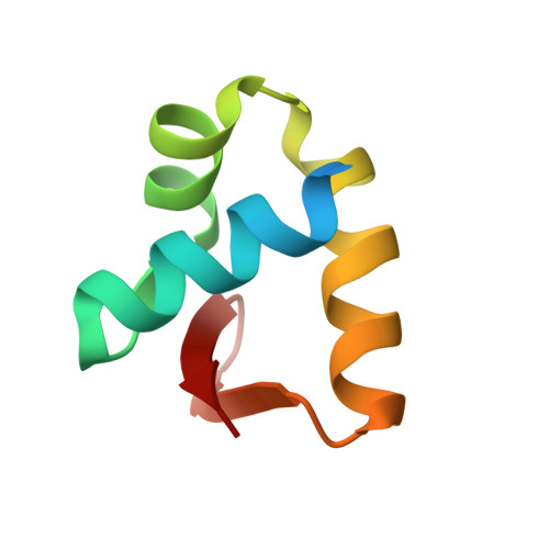
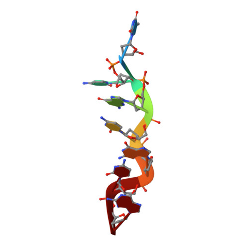
Structure of the DLM-1-Z-DNA complex reveals a conserved family of Z-DNA-binding proteins.

Schwartz, T.Behlke, J.Lowenhaupt, K.Heinemann, U.Rich, A.

(2001) Nat Struct Biol 8: 761-765

  • DOI: https://doi.org/10.1038/nsb0901-761
  • Primary Citation of Related Structures:  
    1J75

  • PubMed Abstract: 

    The first crystal structure of a protein, the Z alpha high affinity binding domain of the RNA editing enzyme ADAR1, bound to left-handed Z-DNA was recently described. The essential set of residues determined from this structure to be critical for Z-DNA recognition was used to search the database for other proteins with the potential for Z-DNA binding. We found that the tumor-associated protein DLM-1 contains a domain with remarkable sequence similarities to Z alpha(ADAR). Here we report the crystal structure of this DLM-1 domain bound to left-handed Z-DNA at 1.85 A resolution. Comparison of Z-DNA binding by DLM-1 and ADAR1 reveals a common structure-specific recognition core within the binding domain. However, the domains differ in certain residues peripheral to the protein-DNA interface. These structures reveal a general mechanism of Z-DNA recognition, suggesting the existence of a family of winged-helix proteins sharing a common Z-DNA binding motif.


  • Organizational Affiliation

    Max-Delbrück-Centrum für Molekulare Medizin, Robert-Rössle-Str. 10, D-13125 Berlin, Germany. Thomas.Schwartz@mail.rockefeller.edu


Macromolecules

Find similar proteins by:  (by identity cutoff)  |  3D Structure
Entity ID: 2
MoleculeChains Sequence LengthOrganismDetailsImage
Tumor Stroma and Activated Macrophage Protein DLM-1B [auth A]67Mus musculusMutation(s): 0 
UniProt
Find proteins for Q9QY24 (Mus musculus)
Explore Q9QY24 
Go to UniProtKB:  Q9QY24
Entity Groups  
Sequence Clusters30% Identity50% Identity70% Identity90% Identity95% Identity100% Identity
UniProt GroupQ9QY24
Sequence Annotations
Expand
  • Reference Sequence

Find similar nucleic acids by:  Sequence   |   3D Structure  

Entity ID: 1
MoleculeChains LengthOrganismImage
5'-D(*TP*CP*GP*CP*GP*CP*G)-3'A [auth B]7N/A
Sequence Annotations
Expand
  • Reference Sequence
Experimental Data & Validation

Experimental Data

  • Method: X-RAY DIFFRACTION
  • Resolution: 1.85 Å
  • R-Value Free: 0.240 
  • R-Value Work: 0.218 
  • R-Value Observed: 0.225 
  • Space Group: P 65 2 2
Unit Cell:
Length ( Å )Angle ( ˚ )
a = 63.611α = 90
b = 63.611β = 90
c = 72.238γ = 120
Software Package:
Software NamePurpose
EPMRphasing
CNSrefinement
DENZOdata reduction
SCALEPACKdata scaling

Structure Validation

View Full Validation Report


Entry History 

Deposition Data

Revision History  (Full details and data files)

  • Version 1.0: 2001-09-01
    Type: Initial release
  • Version 1.1: 2008-04-27
    Changes: Version format compliance
  • Version 1.2: 2011-07-13
    Changes: Version format compliance
  • Version 1.3: 2011-11-16
    Changes: Atomic model
  • Version 1.4: 2023-08-16
    Changes: Advisory, Data collection, Database references, Refinement description