1IYX

Crystal structure of enolase from Enterococcus hirae


Experimental Data Snapshot

  • Method: X-RAY DIFFRACTION
  • Resolution: 2.80 Å
  • R-Value Free: 0.239 
  • R-Value Work: 0.175 
  • R-Value Observed: 0.246 

wwPDB Validation   3D Report Full Report


This is version 1.3 of the entry. See complete history


Literature

Crystal Structure of Enterococcus hirae Enolase at 2.8 A Resolution

Hosaka, T.Meguro, T.Yamato, I.Shirakihara, Y.

(2003) J Biochem 133: 817-823

  • DOI: https://doi.org/10.1093/jb/mvg104
  • Primary Citation of Related Structures:  
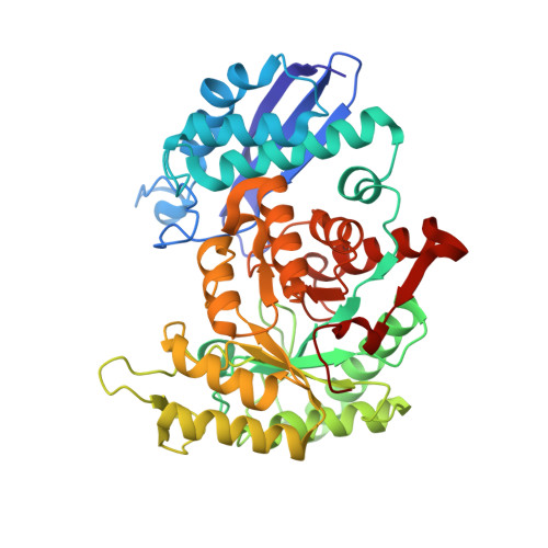
    1IYX

  • PubMed Abstract: 

    We report the crystal structure of an enolase from Enterococcus hirae, which is the first report of a structure determination among gram-positive bacteria. We isolated the enolase gene and determined the base sequence. The amino acid sequence deduced from the DNA sequence suggests that this enolase is composed of 431 amino acids. The amino acid sequence is very similar to those of enolases from eukaryotic and prokaryotic organisms, being 65% and 50% identical to enolases from Escherichia coli and yeast, respectively. The enolase prepared from E. hirae lysate yielded crystals containing one dimer per asymmetric unit. X-ray diffraction patterns were obtained at 2.8 A resolution on a SPring-8 synchrotron radiation source. Crystals belong to space group I4 with unit cell dimensions of a = b = 153.5 A, c = 90.7 A. The E. hirae, yeast, E. coli and lobster enolase structures are very similar. The E. hirae enolase takes an "Open" conformation. The regions in the structure that differ most from other enolases are loops L4 (132-140) and L3 (244-265). Considering the positions of these loops relative to the active site, they seem to have no direct involvement in function. Our findings show that the three dimensional structure of an important enzyme in the glycolytic pathway is evolutionarily conserved among eukaryotes and prokaryotes, including gram-positive bacteria.


  • Organizational Affiliation

    Department of Biological Science and Technology, Tokyo University of Science, 2641 Yamazaki, Noda-shi, Chiba 278-8510.


Macromolecules
Find similar proteins by:  (by identity cutoff)  |  3D Structure
Entity ID: 1
MoleculeChains Sequence LengthOrganismDetailsImage
ENOLASE
A, B
432Enterococcus hiraeMutation(s): 0 
EC: 4.2.1.11
UniProt
Find proteins for Q8GR70 (Enterococcus hirae)
Explore Q8GR70 
Go to UniProtKB:  Q8GR70
Entity Groups  
Sequence Clusters30% Identity50% Identity70% Identity90% Identity95% Identity100% Identity
UniProt GroupQ8GR70
Sequence Annotations
Expand
  • Reference Sequence
Small Molecules
Ligands 3 Unique
IDChains Name / Formula / InChI Key2D Diagram3D Interactions
SO4
Query on SO4

Download Ideal Coordinates CCD File 
D [auth A],
J [auth B]
SULFATE ION
O4 S
QAOWNCQODCNURD-UHFFFAOYSA-L
GOL
Query on GOL

Download Ideal Coordinates CCD File 
E [auth A]
F [auth A]
G [auth A]
H [auth A]
K [auth B]
E [auth A],
F [auth A],
G [auth A],
H [auth A],
K [auth B],
L [auth B]
GLYCEROL
C3 H8 O3
PEDCQBHIVMGVHV-UHFFFAOYSA-N
MG
Query on MG

Download Ideal Coordinates CCD File 
C [auth A],
I [auth B]
MAGNESIUM ION
Mg
JLVVSXFLKOJNIY-UHFFFAOYSA-N
Experimental Data & Validation

Experimental Data

  • Method: X-RAY DIFFRACTION
  • Resolution: 2.80 Å
  • R-Value Free: 0.239 
  • R-Value Work: 0.175 
  • R-Value Observed: 0.246 
  • Space Group: I 4
Unit Cell:
Length ( Å )Angle ( ˚ )
a = 153.51α = 90
b = 153.51β = 90
c = 90.66γ = 90
Software Package:
Software NamePurpose
MOSFLMdata reduction
SCALAdata scaling
MOLREPphasing
CNSrefinement
CCP4data scaling

Structure Validation

View Full Validation Report


Entry History 

Deposition Data

Revision History  (Full details and data files)

  • Version 1.0: 2003-07-29
    Type: Initial release
  • Version 1.1: 2008-04-27
    Changes: Version format compliance
  • Version 1.2: 2011-07-13
    Changes: Non-polymer description, Version format compliance
  • Version 1.3: 2023-10-25
    Changes: Data collection, Database references, Derived calculations, Refinement description