1IS6

MES-Liganded Congerin II


Experimental Data Snapshot

  • Method: X-RAY DIFFRACTION
  • Resolution: 1.70 Å
  • R-Value Free: 0.232 
  • R-Value Work: 0.187 
  • R-Value Observed: 0.188 

wwPDB Validation   3D Report Full Report


This is version 2.1 of the entry. See complete history


Literature

Crystal structure of a conger eel galectin (congerin II) at 1.45 A resolution: Implication for the accelerated evolution of a new ligand-binding site following gene duplication

Shirai, T.Matsui, Y.Shionyu-Mitsuyama, C.Yamane, T.Kamiya, H.Ishii, C.Ogawa, T.Muramoto, K.

(2002) J Mol Biol 321: 879-889

  • DOI: https://doi.org/10.1016/s0022-2836(02)00700-3
  • Primary Citation of Related Structures:  
    1IS5, 1IS6

  • PubMed Abstract: 

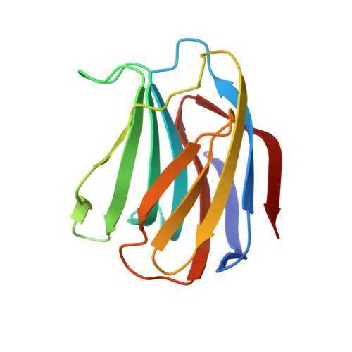
    The crystal structure of congerin II, a galectin family lectin from conger eel, was determined at 1.45A resolution. The previously determined structure of its isoform, congerin I, had revealed a fold evolution via strand swap; however, the structure of congerin II described here resembles other prototype galectins. A comparison of the two congerin genes with that of several other galectins suggests acceralated evolution of both congerin genes following gene duplication. The presence of a Mes (2-[N-morpholino]ethanesulfonic acid) molecule near the carbohydrate-binding site in the crystal structure points to the possibility of an additional binding site in congerin II. The binding site consists of a group of residues that had been replaced following gene duplication suggesting that the binding site was built under selective pressure. Congerin II may be a protein specialized for biological defense with an affinity for target carbohydrates on parasites' cell surface.


  • Organizational Affiliation

    Department of Biotechnology and Biomaterial Chemistry, Graduate School of Engineering, Nagoya University, Chikusa-Ku, Japan. i45282a@nucc.cc.nagoya-u.ac.jp


Macromolecules
Find similar proteins by:  (by identity cutoff)  |  3D Structure
Entity ID: 1
MoleculeChains Sequence LengthOrganismDetailsImage
Congerin II135Conger myriasterMutation(s): 0 
UniProt
Find proteins for Q9YIC2 (Conger myriaster)
Explore Q9YIC2 
Go to UniProtKB:  Q9YIC2
Entity Groups  
Sequence Clusters30% Identity50% Identity70% Identity90% Identity95% Identity100% Identity
UniProt GroupQ9YIC2
Sequence Annotations
Expand
  • Reference Sequence
Small Molecules
Ligands 1 Unique
IDChains Name / Formula / InChI Key2D Diagram3D Interactions
MES
Query on MES

Download Ideal Coordinates CCD File 
B [auth A]2-(N-MORPHOLINO)-ETHANESULFONIC ACID
C6 H13 N O4 S
SXGZJKUKBWWHRA-UHFFFAOYSA-N
Experimental Data & Validation

Experimental Data

  • Method: X-RAY DIFFRACTION
  • Resolution: 1.70 Å
  • R-Value Free: 0.232 
  • R-Value Work: 0.187 
  • R-Value Observed: 0.188 
  • Space Group: P 42 21 2
Unit Cell:
Length ( Å )Angle ( ˚ )
a = 61.4α = 90
b = 61.4β = 90
c = 80.8γ = 90
Software Package:
Software NamePurpose
AMoREphasing
X-PLORrefinement
DENZOdata reduction
SCALEPACKdata scaling

Structure Validation

View Full Validation Report


Entry History 

Deposition Data

Revision History  (Full details and data files)

  • Version 1.0: 2002-09-18
    Type: Initial release
  • Version 1.1: 2008-04-27
    Changes: Version format compliance
  • Version 1.2: 2011-07-13
    Changes: Version format compliance
  • Version 2.0: 2023-12-27
    Changes: Atomic model, Data collection, Database references, Derived calculations
  • Version 2.1: 2024-04-03
    Changes: Advisory, Derived calculations, Refinement description