Cleavage of the iron-methionine bond in c-type cytochromes: crystal structure of oxidized and reduced cytochrome c(2) from Rhodopseudomonas palustris and its ammonia complex.
Geremia, S., Garau, G., Vaccari, L., Sgarra, R., Viezzoli, M.S., Calligaris, M., Randaccio, L.(2002) Protein Sci 11: 6-17
- PubMed: 11742117
- DOI: https://doi.org/10.1110/ps.ps.13102
- Primary Citation of Related Structures:
1FJ0, 1I8O, 1I8P - PubMed Abstract:
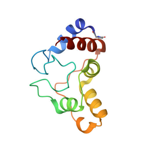
The three-dimensional structures of the native cytochrome c(2) from Rhodopseudomonas palustris and of its ammonia complex have been obtained at pH 4.4 and pH 8.5, respectively. The structure of the native form has been refined in the oxidized state at 1.70 A and in the reduced state at 1.95 A resolution. These are the first high-resolution crystal structures in both oxidation states of a cytochrome c(2) with relatively high redox potential (+350 mV). The differences between the two oxidation states of the native form, including the position of internal water molecules, are small. The unusual six-residue insertion Gly82-Ala87, which precedes the heme binding Met93, forms an isolated 3(10)-helix secondary structural element not previously observed in other c-type cytochromes. Furthermore, this cytochrome shows an external methionine residue involved in a strained folding near the exposed edge of the heme. The structural comparison of the present cytochrome c(2) with other c-type cytochromes has revealed that the presence of such a residue, with torsion angles phi and psi of approximately -140 and -130 degrees, respectively, is a typical feature of this family of proteins. The refined crystal structure of the ammonia complex, obtained at 1.15 A resolution, shows that the sulphur atom of the Met93 axial ligand does not coordinate the heme iron atom, but is replaced by an exogenous ammonia molecule. This is the only example so far reported of an X-ray structure with the heme iron coordinated by an ammonia molecule. The detachment of Met93 is accompanied by a very localized change in backbone conformation, involving mainly the residues Lys92, Met93, and Thr94. Previous studies under typical denaturing conditions, including high-pH values and the presence of exogenous ligands, have shown that the detachment of the Met axial ligand is a basic step in the folding/unfolding process of c-type cytochromes. The ammonia adduct represents a structural model for this important step of the unfolding pathway. Factors proposed to be important for the methionine dissociation are the strength of the H-bond between the Met93 and Tyr66 residues that stabilizes the native form, and the presence in this bacterial cytochrome c(2) of the rare six-residue insertion in the helix 3(10) conformation that increases Met loop flexibility.
Organizational Affiliation:
Centro di Eccellenza di Biocristallografia, Dipartimento di Scienze Chimiche, Università di Trieste, I-34127 Trieste, Italy. geremia@univ.trieste.it