1I3K

MOLECULAR BASIS FOR SEVERE EPIMERASE-DEFICIENCY GALACTOSEMIA: X-RAY STRUCTURE OF THE HUMAN V94M-SUBSTITUTED UDP-GALACTOSE 4-EPIMERASE


Experimental Data Snapshot

  • Method: X-RAY DIFFRACTION
  • Resolution: 1.50 Å
  • R-Value Free: 0.198 
  • R-Value Work: 0.174 
  • R-Value Observed: 0.175 

wwPDB Validation   3D Report Full Report


This is version 1.5 of the entry. See complete history


Literature

Molecular basis for severe epimerase deficiency galactosemia. X-ray structure of the human V94m-substituted UDP-galactose 4-epimerase.

Thoden, J.B.Wohlers, T.M.Fridovich-Keil, J.L.Holden, H.M.

(2001) J Biol Chem 276: 20617-20623

  • DOI: https://doi.org/10.1074/jbc.M101304200
  • Primary Citation of Related Structures:  
    1I3K, 1I3L, 1I3M, 1I3N

  • PubMed Abstract: 

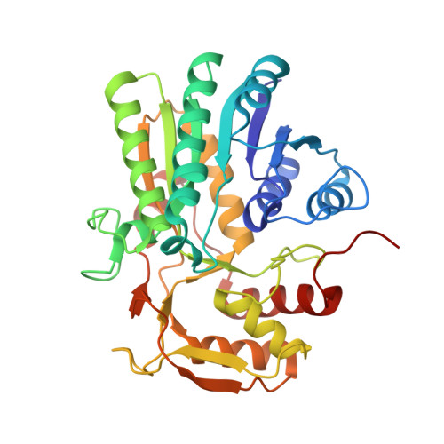
    Galactosemia is an inherited disorder characterized by an inability to metabolize galactose. Although classical galactosemia results from impairment of the second enzyme of the Leloir pathway, namely galactose-1-phosphate uridylyltransferase, alternate forms of the disorder can occur due to either galactokinase or UDP-galactose 4-epimerase deficiencies. One of the more severe cases of epimerase deficiency galactosemia arises from an amino acid substitution at position 94. It has been previously demonstrated that the V94M protein is impaired relative to the wild-type enzyme predominantly at the level of V(max) rather than K(m). To address the molecular consequences the mutation imparts on the three-dimensional architecture of the enzyme, we have solved the structures of the V94M-substituted human epimerase complexed with NADH and UDP-glucose, UDP-galactose, UDP-GlcNAc, or UDP-GalNAc. In the wild-type enzyme, the hydrophobic side chain of Val(94) packs near the aromatic group of the catalytic Tyr(157) and serves as a molecular "fence" to limit the rotation of the glycosyl portions of the UDP-sugar substrates within the active site. The net effect of the V94M substitution is an opening up of the Ala(93) to Glu(96) surface loop, which allows free rotation of the sugars into nonproductive binding modes.


  • Organizational Affiliation

    Department of Biochemistry, University of Wisconsin, Madison, Wisconsin, 53706, USA.


Macromolecules
Find similar proteins by:  (by identity cutoff)  |  3D Structure
Entity ID: 1
MoleculeChains Sequence LengthOrganismDetailsImage
UDP-GLUCOSE 4-EPIMERASE
A, B
348Homo sapiensMutation(s): 1 
Gene Names: HGALE
EC: 5.1.3.2
UniProt & NIH Common Fund Data Resources
Find proteins for Q14376 (Homo sapiens)
Explore Q14376 
Go to UniProtKB:  Q14376
PHAROS:  Q14376
GTEx:  ENSG00000117308 
Entity Groups  
Sequence Clusters30% Identity50% Identity70% Identity90% Identity95% Identity100% Identity
UniProt GroupQ14376
Sequence Annotations
Expand
  • Reference Sequence
Small Molecules
Ligands 5 Unique
IDChains Name / Formula / InChI Key2D Diagram3D Interactions
NAD
Query on NAD

Download Ideal Coordinates CCD File 
E [auth A],
K [auth B]
NICOTINAMIDE-ADENINE-DINUCLEOTIDE
C21 H27 N7 O14 P2
BAWFJGJZGIEFAR-NNYOXOHSSA-N
UPG
Query on UPG

Download Ideal Coordinates CCD File 
F [auth A],
L [auth B]
URIDINE-5'-DIPHOSPHATE-GLUCOSE
C15 H24 N2 O17 P2
HSCJRCZFDFQWRP-JZMIEXBBSA-N
EDO
Query on EDO

Download Ideal Coordinates CCD File 
G [auth A],
M [auth B],
N [auth B]
1,2-ETHANEDIOL
C2 H6 O2
LYCAIKOWRPUZTN-UHFFFAOYSA-N
CL
Query on CL

Download Ideal Coordinates CCD File 
C [auth A],
H [auth B],
I [auth B],
J [auth B]
CHLORIDE ION
Cl
VEXZGXHMUGYJMC-UHFFFAOYSA-M
MG
Query on MG

Download Ideal Coordinates CCD File 
D [auth A]MAGNESIUM ION
Mg
JLVVSXFLKOJNIY-UHFFFAOYSA-N
Experimental Data & Validation

Experimental Data

  • Method: X-RAY DIFFRACTION
  • Resolution: 1.50 Å
  • R-Value Free: 0.198 
  • R-Value Work: 0.174 
  • R-Value Observed: 0.175 
  • Space Group: P 21 21 21
Unit Cell:
Length ( Å )Angle ( ˚ )
a = 78.1α = 90
b = 89.9β = 90
c = 96.9γ = 90
Software Package:
Software NamePurpose
TNTrefinement
d*TREKdata reduction
HKL-2000data scaling
TNTphasing

Structure Validation

View Full Validation Report


Entry History 

Deposition Data

Revision History  (Full details and data files)

  • Version 1.0: 2001-06-20
    Type: Initial release
  • Version 1.1: 2008-04-27
    Changes: Version format compliance
  • Version 1.2: 2011-07-13
    Changes: Version format compliance
  • Version 1.3: 2019-11-20
    Changes: Derived calculations
  • Version 1.4: 2021-10-27
    Changes: Database references, Derived calculations
  • Version 1.5: 2023-08-09
    Changes: Data collection, Refinement description