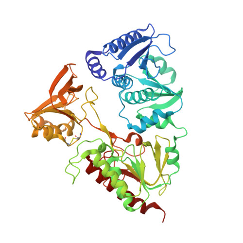
Structure of intact AhpF reveals a mirrored thioredoxin-like active site and implies large domain rotations during catalysis.
Wood, Z.A., Poole, L.B., Karplus, P.A.(2001) Biochemistry 40: 3900-3911
- PubMed: 11300769
- DOI: https://doi.org/10.1021/bi002765p
- Primary Citation of Related Structures:
1HYU - PubMed Abstract:
AhpF, a homodimer of 57 kDa subunits, is a flavoenzyme which catalyzes the NADH-dependent reduction of redox-active disulfide bonds in the peroxidase AhpC, a member of the recently identified peroxiredoxin class of antioxidant enzymes. The structure of AhpF from Salmonella typhimurium at 2.0 A resolution, determined using multiwavelength anomalous dispersion, shows that the C-terminal portion of AhpF (residues 210-521) is structurally like Escherichia coli thioredoxin reductase. In addition, AhpF has an N-terminal domain (residues 1-196) formed from two contiguous thioredoxin folds, but containing just a single redox-active disulfide (Cys129-Cys132). A flexible linker (residues 197-209) connects the domains, consistent with experiments showing that the N-terminal domain acts as an appended substrate, first being reduced by the C-terminal portion of AhpF, and subsequently reducing AhpC. Modeling studies imply that an intrasubunit electron transfer accounts for the reduction of the N-terminal domain in dimeric AhpF. Furthermore, comparing the N-terminal domain with protein disulfide oxidoreductase from Pyrococcus furiosis, we describe a new class of protein disulfide oxidoreductases based on a novel mirror-image active site arrangement, with a distinct carboxylate (Glu86) being functionally equivalent to the key acid (Asp26) of E. coli thioredoxin. A final fortuitous result is that the N-terminal redox center is reduced and provides a high-resolution view of the thiol-thiolate hydrogen bond that has been predicted to stabilize the attacking thiolate in thioredoxin-like proteins.
Organizational Affiliation:
Department of Biochemistry and Biophysics, Oregon State University, Corvallis, Oregon 97331, USA.