1HQH

CRYSTAL STRUCTURE OF THE BINUCLEAR MANGANESE METALLOENZYME ARGINASE COMPLEXED WITH NOR-N-HYDROXY-L-ARGININE


Experimental Data Snapshot

  • Method: X-RAY DIFFRACTION
  • Resolution: 2.80 Å
  • R-Value Free: 0.259 
  • R-Value Work: 0.286 
  • R-Value Observed: 0.286 

wwPDB Validation   3D Report Full Report


This is version 1.3 of the entry. See complete history


Literature

Mechanistic and metabolic inferences from the binding of substrate analogues and products to arginase.

Cox, J.D.Cama, E.Colleluori, D.M.Pethe, S.Boucher, J.L.Mansuy, D.Ash, D.E.Christianson, D.W.

(2001) Biochemistry 40: 2689-26701

  • DOI: https://doi.org/10.1021/bi002318+
  • Primary Citation of Related Structures:  
    1HQF, 1HQG, 1HQH

  • PubMed Abstract: 

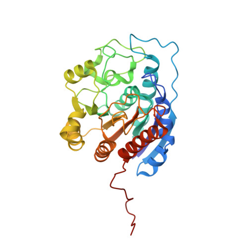
    Arginase is a binuclear Mn(2+) metalloenzyme that catalyzes the hydrolysis of L-arginine to L-ornithine and urea. X-ray crystal structures of arginase complexed to substrate analogues N(omega)-hydroxy-L-arginine and N(omega)-hydroxy-nor-L-arginine, as well as the products L-ornithine and urea, complete a set of structural "snapshots" along the reaction coordinate of arginase catalysis when interpreted along with the X-ray crystal structure of the arginase-transition-state analogue complex described in Kim et al. [Kim, N. N., Cox, J. D., Baggio, R. F., Emig, F. A., Mistry, S., Harper, S. L., Speicher, D. W., Morris, Jr., S. M., Ash, D. E., Traish, A. M., and Christianson, D. W. (2001) Biochemistry 40, 2678-2688]. Taken together, these structures render important insight on the structural determinants of tight binding inhibitors. Furthermore, we demonstrate for the first time the structural mechanistic link between arginase and NO synthase through their respective complexes with N(omega)-hydroxy-L-arginine. That N(omega)-hydroxy-L-arginine is a catalytic intermediate for NO synthase and an inhibitor of arginase reflects the reciprocal metabolic relationship between these two critical enzymes of L-arginine catabolism.


  • Organizational Affiliation

    Roy and Diana Vagelos Laboratories, Department of Chemistry, University of Pennsylvania, Philadelphia, Pennsylvania 19104-6323, USA.


Macromolecules
Find similar proteins by:  (by identity cutoff)  |  3D Structure
Entity ID: 1
MoleculeChains Sequence LengthOrganismDetailsImage
ARGINASE 1
A, B, C
323Rattus norvegicusMutation(s): 0 
EC: 3.5.3.1
UniProt
Find proteins for P07824 (Rattus norvegicus)
Explore P07824 
Go to UniProtKB:  P07824
Entity Groups  
Sequence Clusters30% Identity50% Identity70% Identity90% Identity95% Identity100% Identity
UniProt GroupP07824
Sequence Annotations
Expand
  • Reference Sequence
Small Molecules
Binding Affinity Annotations 
IDSourceBinding Affinity
NNH PDBBind:  1HQH Ki: 500 (nM) from 1 assay(s)
BindingDB:  1HQH Kd: min: 47, max: 517 (nM) from 2 assay(s)
IC50: min: 800, max: 6000 (nM) from 5 assay(s)
Experimental Data & Validation

Experimental Data

  • Method: X-RAY DIFFRACTION
  • Resolution: 2.80 Å
  • R-Value Free: 0.259 
  • R-Value Work: 0.286 
  • R-Value Observed: 0.286 
  • Space Group: P 32
Unit Cell:
Length ( Å )Angle ( ˚ )
a = 88α = 90
b = 88β = 90
c = 112.4γ = 120
Software Package:
Software NamePurpose
AMoREphasing
CNSrefinement
DENZOdata reduction
SCALEPACKdata scaling

Structure Validation

View Full Validation Report


Entry History 

Deposition Data

Revision History  (Full details and data files)

  • Version 1.0: 2001-04-04
    Type: Initial release
  • Version 1.1: 2008-04-27
    Changes: Version format compliance
  • Version 1.2: 2011-07-13
    Changes: Version format compliance
  • Version 1.3: 2024-02-07
    Changes: Data collection, Database references, Derived calculations