1HDM

HISTOCOMPATIBILITY ANTIGEN HLA-DM


Experimental Data Snapshot

  • Method: X-RAY DIFFRACTION
  • Resolution: 2.50 Å
  • R-Value Free: 0.257 
  • R-Value Work: 0.197 
  • R-Value Observed: 0.197 

wwPDB Validation   3D Report Full Report


This is version 1.3 of the entry. See complete history


Literature

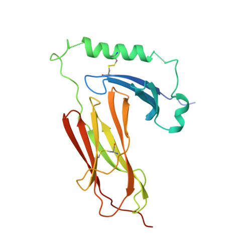
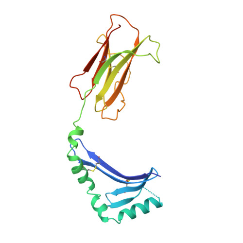
The structure of HLA-DM, the peptide exchange catalyst that loads antigen onto class II MHC molecules during antigen presentation.

Mosyak, L.Zaller, D.M.Wiley, D.C.

(1998) Immunity 9: 377-383

  • DOI: https://doi.org/10.1016/s1074-7613(00)80620-2
  • Primary Citation of Related Structures:  
    1HDM

  • PubMed Abstract: 

    The three-dimensional structure of the soluble ecto-domain of HLA-DM has been determined to 2.5 A resolution by X-ray crystallography. HLA-DM has both peptide exchange activity and acts as a chaperone to peptide-free class II MHC molecules. As predicted, the structure is similar to that of classical class II MHC molecules except that the peptide-binding site is altered to an almost fully closed groove. An unusual cavity is found at the center of the region that binds peptides in class II MHC molecules, and a tryptophanrich lateral surface is identified that is a candidate both for binding to HLA-DR, to effect catalysis, and to HLA-DO, an inhibitor.


  • Organizational Affiliation

    Department of Cellular and Molecular Biology, Howard Hughes Medical Institute, Harvard University, Cambridge, Massachusetts 02138, USA. mosyak@crystal.harvard.edu


Macromolecules
Find similar proteins by:  (by identity cutoff)  |  3D Structure
Entity ID: 1
MoleculeChains Sequence LengthOrganismDetailsImage
PROTEIN (CLASS II HISTOCOMPATIBILITY ANTIGEN, M ALPHA CHAIN)201Homo sapiensMutation(s): 0 
UniProt & NIH Common Fund Data Resources
Find proteins for P28067 (Homo sapiens)
Explore P28067 
Go to UniProtKB:  P28067
PHAROS:  P28067
GTEx:  ENSG00000204257 
Entity Groups  
Sequence Clusters30% Identity50% Identity70% Identity90% Identity95% Identity100% Identity
UniProt GroupP28067
Sequence Annotations
Expand
  • Reference Sequence
Find similar proteins by:  (by identity cutoff)  |  3D Structure
Entity ID: 2
MoleculeChains Sequence LengthOrganismDetailsImage
PROTEIN (CLASS II HISTOCOMPATIBILITY ANTIGEN, M BETA CHAIN)193Homo sapiensMutation(s): 0 
UniProt & NIH Common Fund Data Resources
Find proteins for P28068 (Homo sapiens)
Explore P28068 
Go to UniProtKB:  P28068
PHAROS:  P28068
GTEx:  ENSG00000242574 
Entity Groups  
Sequence Clusters30% Identity50% Identity70% Identity90% Identity95% Identity100% Identity
UniProt GroupP28068
Sequence Annotations
Expand
  • Reference Sequence
Experimental Data & Validation

Experimental Data

  • Method: X-RAY DIFFRACTION
  • Resolution: 2.50 Å
  • R-Value Free: 0.257 
  • R-Value Work: 0.197 
  • R-Value Observed: 0.197 
  • Space Group: C 2 2 21
Unit Cell:
Length ( Å )Angle ( ˚ )
a = 95.39α = 90
b = 109.93β = 90
c = 105.12γ = 90
Software Package:
Software NamePurpose
DENZOdata reduction
SCALEPACKdata scaling
AMoREphasing
X-PLORrefinement

Structure Validation

View Full Validation Report


Entry History 

Deposition Data

Revision History  (Full details and data files)

  • Version 1.0: 1999-09-10
    Type: Initial release
  • Version 1.1: 2008-04-27
    Changes: Version format compliance
  • Version 1.2: 2011-07-13
    Changes: Version format compliance
  • Version 1.3: 2023-08-09
    Changes: Data collection, Database references, Refinement description