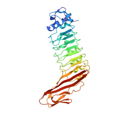
1H6T

Internalin B: crystal structure of fused N-terminal domains.


Experimental Data Snapshot

  • Method: X-RAY DIFFRACTION
  • Resolution: 1.60 Å
  • R-Value Free: 0.197 
  • R-Value Work: 0.165 

wwPDB Validation   3D Report Full Report


This is version 1.4 of the entry. See complete history


Literature

Internalins from the human pathogen Listeria monocytogenes combine three distinct folds into a contiguous internalin domain.

Schubert, W.D.Gobel, G.Diepholz, M.Darji, A.Kloer, D.Hain, T.Chakraborty, T.Wehland, J.Domann, E.Heinz, D.W.

(2001) J Mol Biol 312: 783-794

  • DOI: https://doi.org/10.1006/jmbi.2001.4989
  • Primary Citation of Related Structures:  
    1H6T, 1H6U

  • PubMed Abstract: 

    Listeria monocytogenes is an opportunistic, food-borne human and animal pathogen. Host cell invasion requires the action of the internalins A (InlA) and B (InlB), which are members of a family of listerial cell-surface proteins. Common to these proteins are three distinctive N-terminal domains that have been shown to direct host cell-specific invasion for InlA and InlB. Here, we present the high-resolution crystal structures of these domains present in InlB and InlH, and show that they constitute a single "internalin domain". In this internalin domain, a central LRR region is flanked contiguously by a truncated EF-hand-like cap and an immunoglobulin (Ig)-like fold. The extended beta-sheet, resulting from the distinctive fusion of the LRR and the Ig-like folds, constitutes an adaptable concave interaction surface, which we propose is responsible for the specific recognition of the host cellular binding partners during infection.


  • Organizational Affiliation

    Department of Structural Biology, German Research Center for Biotechnology, Braunschweig.


Macromolecules
Find similar proteins by:  (by identity cutoff)  |  3D Structure
Entity ID: 1
MoleculeChains Sequence LengthOrganismDetailsImage
INTERNALIN B291Listeria monocytogenesMutation(s): 0 
UniProt
Find proteins for P0DQD2 (Listeria monocytogenes serovar 1/2a (strain ATCC BAA-679 / EGD-e))
Explore P0DQD2 
Go to UniProtKB:  P0DQD2
Entity Groups  
Sequence Clusters30% Identity50% Identity70% Identity90% Identity95% Identity100% Identity
UniProt GroupP0DQD2
Sequence Annotations
Expand
  • Reference Sequence
Experimental Data & Validation

Experimental Data

  • Method: X-RAY DIFFRACTION
  • Resolution: 1.60 Å
  • R-Value Free: 0.197 
  • R-Value Work: 0.165 
  • Space Group: P 21 21 21
Unit Cell:
Length ( Å )Angle ( ˚ )
a = 32.953α = 90
b = 72.818β = 90
c = 115.57γ = 90
Software Package:
Software NamePurpose
REFMACrefinement
DENZOdata reduction
SCALEPACKdata scaling
AMoREphasing

Structure Validation

View Full Validation Report


Entry History 

Deposition Data

Revision History  (Full details and data files)

  • Version 1.0: 2001-10-11
    Type: Initial release
  • Version 1.1: 2011-05-08
    Changes: Version format compliance
  • Version 1.2: 2011-07-13
    Changes: Version format compliance
  • Version 1.3: 2019-05-15
    Changes: Data collection, Database references, Experimental preparation, Other
  • Version 1.4: 2023-12-13
    Changes: Data collection, Database references, Refinement description