1H54

Maltose phosphorylase from Lactobacillus brevis


Experimental Data Snapshot

  • Method: X-RAY DIFFRACTION
  • Resolution: 2.15 Å
  • R-Value Free: 0.225 
  • R-Value Work: 0.186 
  • R-Value Observed: 0.186 

wwPDB Validation   3D Report Full Report


This is version 1.1 of the entry. See complete history


Literature

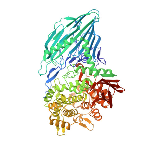
Crystal Structure of Maltose Phosphorylase from Lactobacillus Brevis: Unexpected Evolutionary Relationship with Glucoamylases.

Egloff, M.-P.Uppenberg, J.Haalck, L.Van Tilbeurgh, H.

(2001) Structure 9: 689

  • DOI: https://doi.org/10.1016/s0969-2126(01)00626-8

  • PubMed Abstract: 

    Maltose phosphorylase (MP) is a dimeric enzyme that catalyzes the conversion of maltose and inorganic phosphate into beta-D-glucose-1-phosphate and glucose without requiring any cofactors, such as pyridoxal phosphate. The enzyme is part of operons that are involved in maltose/malto-oligosaccharide metabolism. Maltose phosphorylases have been classified in family 65 of the glycoside hydrolases. No structure is available for any member of this family.


  • Organizational Affiliation

    Architecture et Fonction des Macromolécules Biologiques, Centre National de la Recherche Scientifique, Unite Mixte de Recherche 6098, Université d'Aix-Marseille, I et II, Case 925, 13288, Marseille, France.


Macromolecules
Find similar proteins by:  (by identity cutoff)  |  3D Structure
Entity ID: 1
MoleculeChains Sequence LengthOrganismDetailsImage
MALTOSE PHOSPHORYLASE
A, B
754Levilactobacillus brevisMutation(s): 0 
EC: 2.4.1.8
UniProt
Find proteins for Q7SIE1 (Levilactobacillus brevis)
Explore Q7SIE1 
Go to UniProtKB:  Q7SIE1
Entity Groups  
Sequence Clusters30% Identity50% Identity70% Identity90% Identity95% Identity100% Identity
UniProt GroupQ7SIE1
Sequence Annotations
Expand
  • Reference Sequence
Small Molecules
Ligands 2 Unique
IDChains Name / Formula / InChI Key2D Diagram3D Interactions
PO4
Query on PO4

Download Ideal Coordinates CCD File 
D [auth B]PHOSPHATE ION
O4 P
NBIIXXVUZAFLBC-UHFFFAOYSA-K
K
Query on K

Download Ideal Coordinates CCD File 
C [auth A],
E [auth B]
POTASSIUM ION
K
NPYPAHLBTDXSSS-UHFFFAOYSA-N
Experimental Data & Validation

Experimental Data

  • Method: X-RAY DIFFRACTION
  • Resolution: 2.15 Å
  • R-Value Free: 0.225 
  • R-Value Work: 0.186 
  • R-Value Observed: 0.186 
  • Space Group: P 1 21 1
Unit Cell:
Length ( Å )Angle ( ˚ )
a = 83.75α = 90
b = 102.5β = 111.3
c = 114γ = 90
Software Package:
Software NamePurpose
CNSrefinement
DENZOdata reduction
SCALEPACKdata scaling
AMoREphasing

Structure Validation

View Full Validation Report


Entry History 

Deposition Data

Revision History  (Full details and data files)

  • Version 1.0: 2001-09-05
    Type: Initial release
  • Version 1.1: 2012-06-13
    Changes: Atomic model, Database references, Derived calculations, Non-polymer description, Other, Refinement description, Version format compliance