Active Site Analysis of the Potential Antimicrobial Target Aspartate Semialdehyde Dehydrogenase.
Hadfield, A.T., Shammas, C., Kryger, G., Ringe, D., Petsko, G.A., Ouyang, J., Viola, R.E.(2001) Biochemistry 40: 14475
- PubMed: 11724560
- DOI: https://doi.org/10.1021/bi015713o
- Primary Citation of Related Structures:
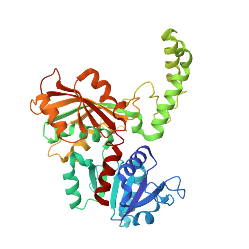
1GL3 - PubMed Abstract:
Aspartate-beta-semialdehyde dehydrogenase (ASADH) lies at the first branch point in the biosynthetic pathway through which bacteria, fungi, and the higher plants synthesize amino acids, including lysine and methionine and the cell wall component diaminopimelate from aspartate. Blocks in this biosynthetic pathway, which is absent in mammals, are lethal, and inhibitors of ASADH may therefore serve as useful antibacterial, fungicidal, or herbicidal agents. We have determined the structure of ASADH from Escherichia coli by crystallography in the presence of its coenzyme and a substrate analogue that acts as a covalent inhibitor. This structure is comparable to that of the covalent intermediate that forms during the reaction catalyzed by ASADH. The key catalytic residues are confirmed as cysteine 135, which is covalently linked to the intermediate during the reaction, and histidine 274, which acts as an acid/base catalyst. The substrate and coenzyme binding residues are also identified, and these active site residues are conserved throughout all of the ASADH sequences. Comparison of the previously determined apo-enzyme structure [Hadfield et al. J. Mol. Biol. (1999) 289, 991-1002] and the complex presented here reveals a conformational change that occurs on binding of NADP that creates a binding site for the amino acid substrate. These results provide a structural explanation for the preferred order of substrate binding that is observed kinetically.
Organizational Affiliation:
Department of Biochemistry, School of Medical Sciences, University of Bristol, University Walk, Bristol BS8 1TD, England. a.t.hadfield@bris.ac.uk