1FXS

GDP-FUCOSE SYNTHETASE FROM ESCHERICHIA COLI COMPLEX WITH NADP


Experimental Data Snapshot

  • Method: X-RAY DIFFRACTION
  • Resolution: 2.30 Å
  • R-Value Observed: 0.163 

wwPDB Validation   3D Report Full Report


Ligand Structure Quality Assessment 


This is version 1.3 of the entry. See complete history


Literature

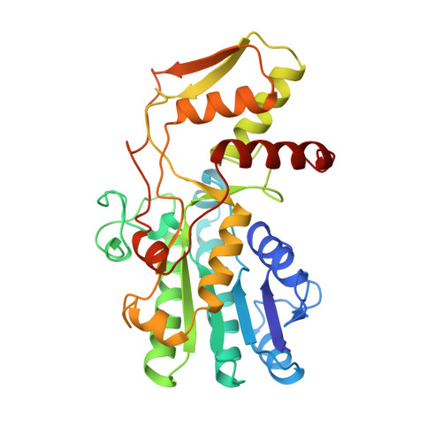
GDP-fucose synthetase from Escherichia coli: structure of a unique member of the short-chain dehydrogenase/reductase family that catalyzes two distinct reactions at the same active site.

Somers, W.S.Stahl, M.L.Sullivan, F.X.

(1998) Structure 6: 1601-1612

  • DOI: https://doi.org/10.1016/s0969-2126(98)00157-9
  • Primary Citation of Related Structures:  
    1BSV, 1FXS, 1GFS

  • PubMed Abstract: 

    . In all species examined, GDP-fucose is synthesized from GDP-mannose in a three-step reaction catalyzed by two enzymes, GDP-mannose 4,6 dehydratase and a dual function 3, 5-epimerase-4-reductase named GDP-fucose synthetase. In this latter aspect fucose biosynthesis differs from that of other deoxy and dideoxy sugars, in which the epimerase and reductase activities are present as separate enzymes. Defects in GDP-fucose biosynthesis have been shown to affect nodulation in bacteria, stem development in plants, and are associated with the immune defect leukocyte adhesion deficiency type II in humans.


  • Organizational Affiliation

    Small Molecule Drug Discovery Genetics Institute, Inc. 87 Cambridgepark Drive, Cambridge, MA 02140, USA.


Macromolecules
Find similar proteins by:  (by identity cutoff)  |  3D Structure
Entity ID: 1
MoleculeChains Sequence LengthOrganismDetailsImage
PROTEIN (GDP-FUCOSE SYNTHETASE)321Escherichia coli K-12Mutation(s): 0 
Gene Names: WCAG
UniProt
Find proteins for P32055 (Escherichia coli (strain K12))
Explore P32055 
Go to UniProtKB:  P32055
Entity Groups  
Sequence Clusters30% Identity50% Identity70% Identity90% Identity95% Identity100% Identity
UniProt GroupP32055
Sequence Annotations
Expand
  • Reference Sequence
Small Molecules
Ligands 1 Unique
IDChains Name / Formula / InChI Key2D Diagram3D Interactions
NAP
Query on NAP

Download Ideal Coordinates CCD File 
B [auth A]NADP NICOTINAMIDE-ADENINE-DINUCLEOTIDE PHOSPHATE
C21 H28 N7 O17 P3
XJLXINKUBYWONI-NNYOXOHSSA-N
Experimental Data & Validation

Experimental Data

  • Method: X-RAY DIFFRACTION
  • Resolution: 2.30 Å
  • R-Value Observed: 0.163 
  • Space Group: P 32 2 1
Unit Cell:
Length ( Å )Angle ( ˚ )
a = 104.2α = 90
b = 104.2β = 90
c = 75.1γ = 120
Software Package:
Software NamePurpose
X-PLORrefinement
DENZOdata reduction
SCALEPACKdata scaling

Structure Validation

View Full Validation Report


Ligand Structure Quality Assessment 


Entry History 

Deposition Data

Revision History  (Full details and data files)

  • Version 1.0: 1999-08-26
    Type: Initial release
  • Version 1.1: 2008-04-27
    Changes: Version format compliance
  • Version 1.2: 2011-07-13
    Changes: Derived calculations, Source and taxonomy, Version format compliance
  • Version 1.3: 2023-08-09
    Changes: Data collection, Database references, Derived calculations, Refinement description