Structure and function of the epidermal growth factor domain of P-selectin.
Freedman, S.J., Sanford, D.G., Bachovchin, W.W., Furie, B.C., Baleja, J.D., Furie, B.(1996) Biochemistry 35: 13733-13744
- PubMed: 8901515
- DOI: https://doi.org/10.1021/bi9610257
- Primary Citation of Related Structures:
1FSB - PubMed Abstract:
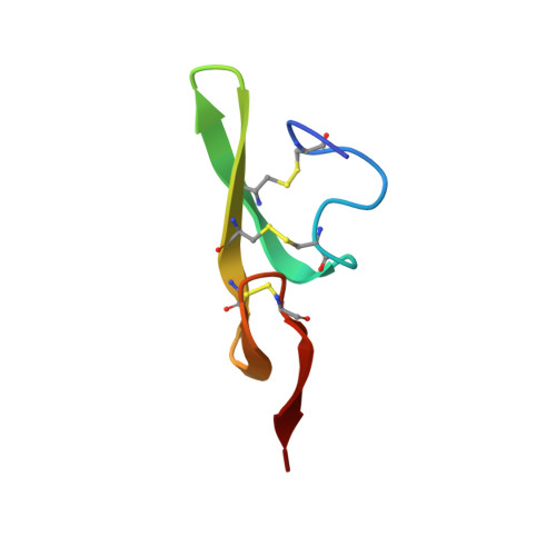
P-selectin is a multidomain adhesion protein on the surface of activated platelets and endothelial cells that functions in the recruitment of leukocytes to the site of inflammation. The amino-terminal lectin and EGF domains constitute the ligand recognition unit. We have produced a synthetic 40-residue P-selectin EGF domain (P-sel:EGF) to examine the structure and function of this domain independent of P-selectin. The peptide was folded in vitro and exhibited the same disulfide bonding pattern as other EGF-like domains. P-sel:EGF did not inhibit P-selectin-mediated cellular adhesion assays, indicating that the lectin domain is also required. We undertook the study of the P-selectin EGF by 1H NMR to determine its structure independent of the lectin domain and to compare its structure to that of E-selectin determined crystallographically [Graves et al. (1994) Nature 367, 532]. Although the binding of P-selectin to its carbohydrate ligand is calcium dependent, and some EGF domains have calcium binding sites, addition of calcium had no effect on the NMR spectrum or on the pH-induced changes. Nearly complete resonance assignments were made from 2D 1H NMR spectra at pH 6.0. Two sections of antiparallel beta-sheet were identified on the basis of the pattern of long-range NOEs, 3JHN alpha coupling constants, and slowly exchanging amides. The solution structure of the peptide backbone was determined using distance geometry and simulated annealing calculations. The backbone RMSD to the geometric average for 19 final structures is 0.64 +/- 0.17 A. The resulting fold closely resembles that of other EGF-like peptides, including the E-selectin EGF domain (RMSD approximately 1.08 A). However, compared to the E-selectin EGF structure which also contains the lectin domain, some residues from 1-11 are less ordered, and novel contacts occur between the amino terminus and the core beta-sheet. Despite marked structural homology of the selectin polypeptide backbones, the selectin EGF surfaces show unique distributions of charged residues, a feature that likely correlates to the functional differences.
Organizational Affiliation:
Center for Hemostasis and Thrombosis Research, New England Medical Center, Boston, Massachusetts, USA.