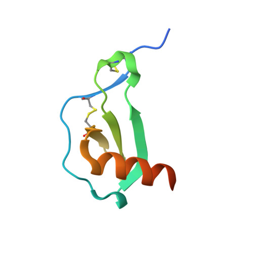
The crystal structure of the chemokine domain of fractalkine shows a novel quaternary arrangement.
Hoover, D.M., Mizoue, L.S., Handel, T.M., Lubkowski, J.(2000) J Biol Chem 275: 23187-23193
- PubMed: 10770945
- DOI: https://doi.org/10.1074/jbc.M002584200
- Primary Citation of Related Structures:
1F2L - PubMed Abstract:
Fractalkine, or neurotactin, is a chemokine that is present in endothelial cells from several tissues, including brain, liver, and kidney. It is the only member of the CX(3)C class of chemokines. Fractalkine contains a chemokine domain (CDF) attached to a membrane-spanning domain via a mucin-like stalk. However, fractalkine can also be proteolytically cleaved from its membrane-spanning domain to release a freely diffusible form. Fractalkine attracts and immobilizes leukocytes by binding to its receptor, CX(3)CR1. The x-ray crystal structure of CDF has been solved and refined to 2.0 A resolution. The CDF monomers form a dimer through an intermolecular beta-sheet. This interaction is somewhat similar to that seen in other dimeric CC chemokine crystal structures. However, the displacement of the first disulfide in CDF causes the dimer to assume a more compact quaternary structure relative to CC chemokines, which is unique to CX(3)C chemokines. Although fractalkine can bind to heparin in vitro, as shown by comparison of electrostatic surface plots with other chemokines and by heparin chromatography, the role of this property in vivo is not well understood.
Organizational Affiliation:
Macromolecular Crystallography Laboratory, Program in Structural Biology, NCI-Frederick Cancer Research and Development Center, Frederick, Maryland 21702, USA.