Structure-Activity Relationships in a Peptidic Alpha7 Nicotinic Acetylcholine Receptor Antagonist
Rogers, J.P., Luginbuhl, P., Pemberton, K., Harty, P., Wemmer, D.E., Stevens, R.C.(2000) J Mol Biol 304: 911
- PubMed: 11124036
- DOI: https://doi.org/10.1006/jmbi.2000.4247
- Primary Citation of Related Structures:
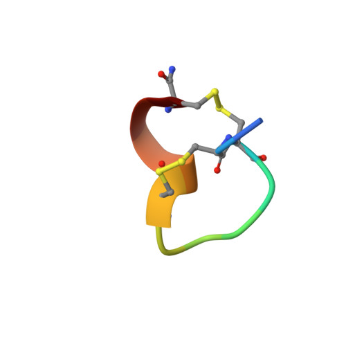
1E74, 1E75, 1E76 - PubMed Abstract:
alpha-Conotoxins are small disulfide-constrained peptide toxins which act as antagonists at specific subtypes of nicotinic acetylcholine receptors (nACh receptors). In this study, we analyzed the structures and activities of three mutants of alpha-conotoxin ImI, a 12 amino acid peptide active at alpha7 nACh receptors, in order to gain insight into the primary and tertiary structural requirements of neuronal alpha-conotoxin specificity. NMR solution structures were determined for mutants R11E, R7L, and D5N, resulting in representative ensembles of 20 conformers with average pairwise RMSD values of 0.46, 0.52, and 0.62 A from their mean structures, respectively, for the backbone atoms N, C(alpha), and C' of residues 2-11. The R11E mutant was found to have activity near that of wild-type ImI, while R7L and D5N demonstrated activities reduced by at least two orders of magnitude. Comparison of the structures reveals a common two-loop architecture, with variations observed in backbone and side-chain dihedral angles as well as surface electrostatic potentials upon mutation. Correlation of these structures and activities with those from previously published studies emphasizes that existing hypotheses regarding the molecular determinants of alpha-conotoxin specificity are not adequate for explaining peptide activity, and suggests that more subtle features, visualized here at the atomic level, are important for receptor binding. These data, in conjunction with reported characterizations of the acetylcholine binding site, support a model of toxin activity in which a single solvent-accessible toxin side-chain anchors the complex, with supporting weak interactions determining both the efficacy and the subtype specificity of the inhibitory activity.
Organizational Affiliation:
Department of Chemistry, University of California, Berkeley, CA, 94720, USA.