1E6V

Methyl-coenzyme M reductase from Methanopyrus kandleri


Experimental Data Snapshot

  • Method: X-RAY DIFFRACTION
  • Resolution: 2.70 Å
  • R-Value Free: 0.278 
  • R-Value Work: 0.239 
  • R-Value Observed: 0.239 

wwPDB Validation   3D Report Full Report


Ligand Structure Quality Assessment 


This is version 1.4 of the entry. See complete history


Literature

Comparison of Three Methyl-Coenzyme M Reductases from Phylogenetically Distant Organisms: Unusual Amino Acid Modification, Conservation and Adaptation

Grabarse, W.Mahlert, F.Shima, S.Thauer, R.K.Ermler, U.

(2000) J Mol Biol 303: 329

  • DOI: https://doi.org/10.1006/jmbi.2000.4136
  • Primary Citation of Related Structures:  
    1E6V, 1E6Y

  • PubMed Abstract: 

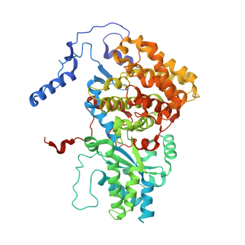
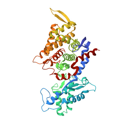
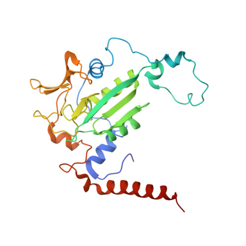
    The nickel enzyme methyl-coenzyme M reductase (MCR) catalyzes the terminal step of methane formation in the energy metabolism of all methanogenic archaea. In this reaction methyl-coenzyme M and coenzyme B are converted to methane and the heterodisulfide of coenzyme M and coenzyme B. The crystal structures of methyl-coenzyme M reductase from Methanosarcina barkeri (growth temperature optimum, 37 degrees C) and Methanopyrus kandleri (growth temperature optimum, 98 degrees C) were determined and compared with the known structure of MCR from Methanobacterium thermoautotrophicum (growth temperature optimum, 65 degrees C). The active sites of MCR from M. barkeri and M. kandleri were almost identical to that of M. thermoautotrophicum and predominantly occupied by coenzyme M and coenzyme B. The electron density at 1.6 A resolution of the M. barkeri enzyme revealed that four of the five modified amino acid residues of MCR from M. thermoautotrophicum, namely a thiopeptide, an S-methylcysteine, a 1-N-methylhistidine and a 5-methylarginine were also present. Analysis of the environment of the unusual amino acid residues near the active site indicates that some of the modifications may be required for the enzyme to be catalytically effective. In M. thermoautotrophicum and M. kandleri high temperature adaptation is coupled with increasing intracellular concentrations of lyotropic salts. This was reflected in a higher fraction of glutamate residues at the protein surface of the thermophilic enzymes adapted to high intracellular salt concentrations.


  • Organizational Affiliation

    Max-Planck-Institut für Biophysik, Heinrich-Hoffmann-Strasse 7, 60528 Frankfurt, Germany.


Macromolecules
Find similar proteins by:  (by identity cutoff)  |  3D Structure
Entity ID: 1
MoleculeChains Sequence LengthOrganismDetailsImage
METHYL-COENZYME M REDUCTASE I ALPHA SUBUNIT
A, D
553Methanopyrus kandleriMutation(s): 0 
UniProt
Find proteins for Q49605 (Methanopyrus kandleri (strain AV19 / DSM 6324 / JCM 9639 / NBRC 100938))
Explore Q49605 
Go to UniProtKB:  Q49605
Entity Groups  
Sequence Clusters30% Identity50% Identity70% Identity90% Identity95% Identity100% Identity
UniProt GroupQ49605
Sequence Annotations
Expand
  • Reference Sequence
Find similar proteins by:  (by identity cutoff)  |  3D Structure
Entity ID: 2
MoleculeChains Sequence LengthOrganismDetailsImage
METHYL-COENZYME M REDUCTASE I BETA SUBUNIT
B, E
443Methanopyrus kandleriMutation(s): 0 
UniProt
Find proteins for Q49601 (Methanopyrus kandleri)
Explore Q49601 
Go to UniProtKB:  Q49601
Entity Groups  
Sequence Clusters30% Identity50% Identity70% Identity90% Identity95% Identity100% Identity
UniProt GroupQ49601
Sequence Annotations
Expand
  • Reference Sequence
Find similar proteins by:  (by identity cutoff)  |  3D Structure
Entity ID: 3
MoleculeChains Sequence LengthOrganismDetailsImage
METHYL-COENZYME M REDUCTASE I GAMMA SUBUNIT
C, F
258Methanopyrus kandleriMutation(s): 0 
UniProt
Find proteins for Q49604 (Methanopyrus kandleri)
Explore Q49604 
Go to UniProtKB:  Q49604
Entity Groups  
Sequence Clusters30% Identity50% Identity70% Identity90% Identity95% Identity100% Identity
UniProt GroupQ49604
Sequence Annotations
Expand
  • Reference Sequence
Experimental Data & Validation

Experimental Data

  • Method: X-RAY DIFFRACTION
  • Resolution: 2.70 Å
  • R-Value Free: 0.278 
  • R-Value Work: 0.239 
  • R-Value Observed: 0.239 
  • Space Group: P 21 21 21
Unit Cell:
Length ( Å )Angle ( ˚ )
a = 80.519α = 90
b = 115.74β = 90
c = 268.51γ = 90
Software Package:
Software NamePurpose
CNSrefinement
DENZOdata reduction
SCALEPACKdata scaling
CNSphasing

Structure Validation

View Full Validation Report


Ligand Structure Quality Assessment 


Entry History 

Deposition Data

Revision History  (Full details and data files)

  • Version 1.0: 2000-10-18
    Type: Initial release
  • Version 1.1: 2015-05-20
    Changes: Database references, Derived calculations, Non-polymer description, Other, Structure summary, Version format compliance
  • Version 1.2: 2017-03-29
    Changes: Non-polymer description
  • Version 1.3: 2019-07-24
    Changes: Advisory, Data collection
  • Version 1.4: 2023-12-13
    Changes: Advisory, Data collection, Database references, Derived calculations, Other, Refinement description