1DWK

STRUCTURE OF CYANASE WITH THE DI-ANION OXALATE BOUND AT THE ENZYME ACTIVE SITE


Experimental Data Snapshot

  • Method: X-RAY DIFFRACTION
  • Resolution: 1.65 Å
  • R-Value Free: 0.181 
  • R-Value Work: 0.146 
  • R-Value Observed: 0.138 

wwPDB Validation   3D Report Full Report


This is version 1.5 of the entry. See complete history


Literature

Structure of Cyanase Reveals that a Novel Dimeric and Decameric Arrangement of Subunits is Required for Formation of the Enzyme Active Site.

Walsh, M.A.Otwinowski, Z.Perrakis, A.Anderson, P.M.Joachimiak, A.

(2000) Structure 8: 505

  • DOI: https://doi.org/10.1016/s0969-2126(00)00134-9
  • Primary Citation of Related Structures:  
    1DW9, 1DWK

  • PubMed Abstract: 

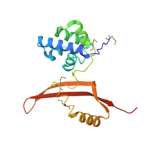
    Cyanase is an enzyme found in bacteria and plants that catalyzes the reaction of cyanate with bicarbonate to produce ammonia and carbon dioxide. In Escherichia coli, cyanase is induced from the cyn operon in response to extracellular cyanate. The enzyme is functionally active as a homodecamer of 17 kDa subunits, and displays half-site binding of substrates or substrate analogs. The enzyme shows no significant amino acid sequence homology with other proteins.


  • Organizational Affiliation

    Biosciences Division/Structural Biology Center, Argonne National Laboratory, Argonne, IL 60439, USA.


Macromolecules
Find similar proteins by:  (by identity cutoff)  |  3D Structure
Entity ID: 1
MoleculeChains Sequence LengthOrganismDetailsImage
CYANATE HYDRATASE
A, B, C, D, E
A, B, C, D, E, F, G, H, I, J
156Escherichia coliMutation(s): 4 
EC: 4.2.1.104
UniProt
Find proteins for P00816 (Escherichia coli (strain K12))
Explore P00816 
Go to UniProtKB:  P00816
Entity Groups  
Sequence Clusters30% Identity50% Identity70% Identity90% Identity95% Identity100% Identity
UniProt GroupP00816
Sequence Annotations
Expand
  • Reference Sequence
Small Molecules
Ligands 2 Unique
IDChains Name / Formula / InChI Key2D Diagram3D Interactions
SO4
Query on SO4

Download Ideal Coordinates CCD File 
AA [auth F]
BA [auth G]
CA [auth G]
DA [auth G]
FA [auth H]
AA [auth F],
BA [auth G],
CA [auth G],
DA [auth G],
FA [auth H],
GA [auth H],
HA [auth I],
IA [auth I],
K [auth A],
KA [auth J],
L [auth A],
LA [auth J],
M [auth A],
N [auth B],
O [auth B],
Q [auth C],
S [auth D],
T [auth D],
U [auth D],
V [auth E],
W [auth E],
X [auth F],
Z [auth F]
SULFATE ION
O4 S
QAOWNCQODCNURD-UHFFFAOYSA-L
OXL
Query on OXL

Download Ideal Coordinates CCD File 
EA [auth G],
JA [auth J],
P [auth B],
R [auth C],
Y [auth F]
OXALATE ION
C2 O4
MUBZPKHOEPUJKR-UHFFFAOYSA-L
Modified Residues  1 Unique
IDChains TypeFormula2D DiagramParent
MSE
Query on MSE
A, B, C, D, E
A, B, C, D, E, F, G, H, I, J
L-PEPTIDE LINKINGC5 H11 N O2 SeMET
Experimental Data & Validation

Experimental Data

  • Method: X-RAY DIFFRACTION
  • Resolution: 1.65 Å
  • R-Value Free: 0.181 
  • R-Value Work: 0.146 
  • R-Value Observed: 0.138 
  • Space Group: P 1
Unit Cell:
Length ( Å )Angle ( ˚ )
a = 76.33α = 70.1
b = 80.93β = 71.95
c = 82.13γ = 66.42
Software Package:
Software NamePurpose
REFMACrefinement
HKL-2000data reduction
HKL-2000data scaling
CCP4phasing

Structure Validation

View Full Validation Report


Entry History 

Deposition Data

Revision History  (Full details and data files)

  • Version 1.0: 2000-05-16
    Type: Initial release
  • Version 1.1: 2011-05-07
    Changes: Version format compliance
  • Version 1.2: 2011-07-13
    Changes: Version format compliance
  • Version 1.3: 2017-07-12
    Changes: Advisory, Derived calculations
  • Version 1.4: 2019-05-08
    Changes: Data collection, Experimental preparation
  • Version 1.5: 2023-12-06
    Changes: Data collection, Database references, Other, Refinement description