Anatomy of a proficient enzyme: the structure of orotidine 5'-monophosphate decarboxylase in the presence and absence of a potential transition state analog.
Miller, B.G., Hassell, A.M., Wolfenden, R., Milburn, M.V., Short, S.A.(2000) Proc Natl Acad Sci U S A 97: 2011-2016
- PubMed: 10681417
- DOI: https://doi.org/10.1073/pnas.030409797
- Primary Citation of Related Structures:
1DQW, 1DQX - PubMed Abstract:
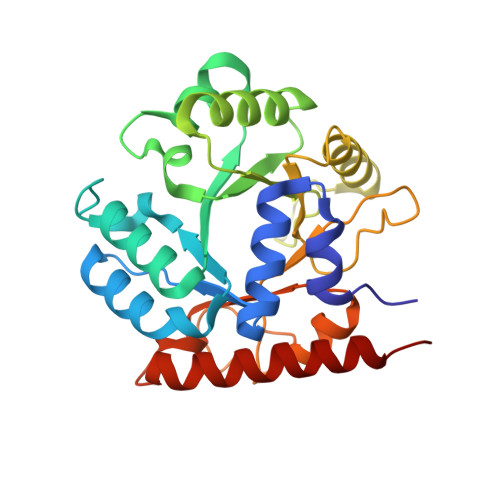
Orotidine 5'-phosphate decarboxylase produces the largest rate enhancement that has been reported for any enzyme. The crystal structure of the recombinant Saccharomyces cerevisiae enzyme has been determined in the absence and presence of the proposed transition state analog 6-hydroxyuridine 5'-phosphate, at a resolution of 2.1 A and 2.4 A, respectively. Orotidine 5'-phosphate decarboxylase folds as a TIM-barrel with the ligand binding site near the open end of the barrel. The binding of 6-hydroxyuridine 5'-phosphate is accompanied by protein loop movements that envelop the ligand almost completely, forming numerous favorable interactions with the phosphoryl group, the ribofuranosyl group, and the pyrimidine ring. Lysine-93 appears to be anchored in such a way as to optimize electrostatic interactions with developing negative charge at C-6 of the pyrimidine ring, and to donate the proton that replaces the carboxylate group at C-6 of the product. In addition, H-bonds from the active site to O-2 and O-4 help to delocalize negative charge in the transition state. Interactions between the enzyme and the phosphoribosyl group anchor the pyrimidine within the active site, helping to explain the phosphoribosyl group's remarkably large contribution to catalysis despite its distance from the site of decarboxylation.
Organizational Affiliation:
Department of Biochemistry, University of North Carolina, Chapel Hill, NC 27599, USA. Research Triangle Park, NC 27709, USA.