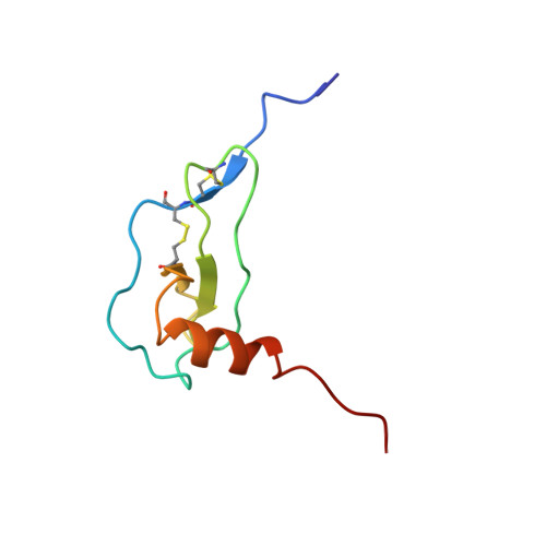
Heteronuclear (1H, 13C, 15N) NMR assignments and solution structure of the monocyte chemoattractant protein-1 (MCP-1) dimer.
Handel, T.M., Domaille, P.J.(1996) Biochemistry 35: 6569-6584
- PubMed: 8639605
- DOI: https://doi.org/10.1021/bi9602270
- Primary Citation of Related Structures:
1DOM, 1DON - PubMed Abstract:
A full high-resolution three-dimensional solution structure of the monocyte chemoattractant protein-1 (MCP-1 or MCAF) homodimer has been determined by heteronuclear multidimensional NMR. MCP-1 is a member of a family of small proteins which play a crucial role in immune surveillance by orchestrating the recruitment of specific leukocytes to areas of immune challenge. The protein was uniformly isotopically enriched with 13C and 15N by expression in Escherichia coli, and complete sequence-specific resonance assignments were obtained by a combination of heteronuclear double- and triple-resonance experiments. The secondary structure was deduced from characteristic patterns of NOEs, 13 C alpha/beta chemical shifts, measurements of 3JHNH alpha scalar couplings, and patterns of slowly exchanging amide protons. Because MCP-1 forms symmetrical homodimers, additional experiments were carried out to unambiguously establish the quaternary contacts. NOEs from these novel experiments were merged with more traditional heteronuclear separated NOE measurements in an iterative strategy to partition the restraints between explicit inter/intrasubunit contacts and a class wherein both were retained as ambiguous. With more than 30 restraints per residue, the three-dimensional structure is well-defined with a backbone rmsd of 0.37 A to the mean over residues 5-69 of the dimer. We compare the structure with those recently reported for the related chemokines MIP-1 beta and RANTES and highlight the differences in terms of receptor specificity and function as well as interpret the known biological activity data of MCP-1 mutants.
Organizational Affiliation:
Department of Molecular and Cell Biology, University of California at Berkeley 94720, USA.