1DE7

INTERACTION OF FACTOR XIII ACTIVATION PEPTIDE WITH ALPHA-THROMBIN: CRYSTAL STRUCTURE OF THE ENZYME-SUBSTRATE COMPLEX


Experimental Data Snapshot

  • Method: X-RAY DIFFRACTION
  • Resolution: 2.00 Å
  • R-Value Free: 0.257 
  • R-Value Work: 0.194 
  • R-Value Observed: 0.194 

wwPDB Validation   3D Report Full Report


This is version 1.3 of the entry. See complete history


Literature

Interaction of the factor XIII activation peptide with alpha -thrombin. Crystal structure of its enzyme-substrate analog complex.

Sadasivan, C.Yee, V.C.

(2000) J Biol Chem 275: 36942-36948

  • DOI: https://doi.org/10.1074/jbc.M006076200
  • Primary Citation of Related Structures:  
    1DE7

  • PubMed Abstract: 

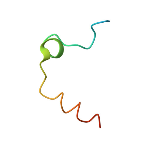
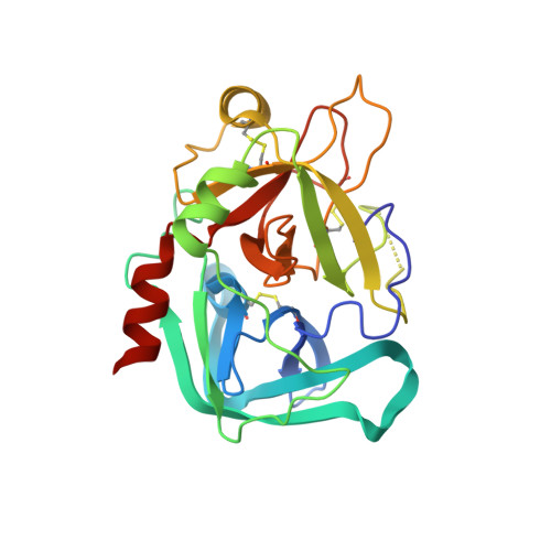
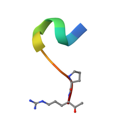
    The serine protease thrombin proteolytically activates blood coagulation factor XIII by cleavage at residue Arg(37); factor XIII in turn cross-links fibrin molecules and gives mechanical stability to the blood clot. The 2.0-A resolution x-ray crystal structure of human alpha-thrombin bound to the factor XIII-(28-37) decapeptide has been determined. This structure reveals the detailed atomic level interactions between the factor XIII activation peptide and thrombin and provides the first high resolution view of this functionally important part of the factor XIII molecule. A comparison of this structure with the crystal structure of fibrinopeptide A complexed with thrombin highlights several important determinants of thrombin substrate interaction. First, the P1 and P2 residues must be compatible with the geometry and chemistry of the S1 and S2 specificity sites in thrombin. Second, a glycine in the P5 position is necessary for the conserved substrate conformation seen in both factor XIII-(28-37) and fibrinopeptide A. Finally, the hydrophobic residues, which occupy the aryl binding site of thrombin determine the substrate conformation further away from the catalytic residues. In the case of factor XIII-(28-37), the aryl binding site is shared by hydrophobic residues P4 (Val(34)) and P9 (Val(29)). A bulkier residue in either of these sites may alter the substrate peptide conformation.


  • Organizational Affiliation

    Department of Molecular Cardiology and Structural Biology Center, Lerner Research Institute, Cleveland Clinic Foundation, Cleveland, Ohio 44195, USA.


Macromolecules
Find similar proteins by:  (by identity cutoff)  |  3D Structure
Entity ID: 1
MoleculeChains Sequence LengthOrganismDetailsImage
ALPHA-THROMBIN (LIGHT CHAIN)A [auth L],
C [auth J]
36Homo sapiensMutation(s): 0 
EC: 3.4.21.5
UniProt & NIH Common Fund Data Resources
Find proteins for P00734 (Homo sapiens)
Explore P00734 
Go to UniProtKB:  P00734
PHAROS:  P00734
GTEx:  ENSG00000180210 
Entity Groups  
Sequence Clusters30% Identity50% Identity70% Identity90% Identity95% Identity100% Identity
UniProt GroupP00734
Sequence Annotations
Expand
  • Reference Sequence
Find similar proteins by:  (by identity cutoff)  |  3D Structure
Entity ID: 2
MoleculeChains Sequence LengthOrganismDetailsImage
ALPHA-THROMBIN (HEAVY CHAIN)B [auth H],
D [auth K]
259Homo sapiensMutation(s): 0 
EC: 3.4.21.5
UniProt & NIH Common Fund Data Resources
Find proteins for P00734 (Homo sapiens)
Explore P00734 
Go to UniProtKB:  P00734
PHAROS:  P00734
GTEx:  ENSG00000180210 
Entity Groups  
Sequence Clusters30% Identity50% Identity70% Identity90% Identity95% Identity100% Identity
UniProt GroupP00734
Sequence Annotations
Expand
  • Reference Sequence

Find similar proteins by:  Sequence   |   3D Structure  

Entity ID: 3
MoleculeChains Sequence LengthOrganismDetailsImage
FACTOR XIII ACTIVATION PEPTIDE (28-37)E [auth A],
F [auth B]
11N/AMutation(s): 0 
Entity Groups  
Sequence Clusters30% Identity50% Identity70% Identity90% Identity95% Identity100% Identity
Sequence Annotations
Expand
  • Reference Sequence
Small Molecules
Modified Residues  1 Unique
IDChains TypeFormula2D DiagramParent
AR7
Query on AR7
E [auth A],
F [auth B]
PEPTIDE-LIKEC6 H17 N4 O2ARG
Experimental Data & Validation

Experimental Data

  • Method: X-RAY DIFFRACTION
  • Resolution: 2.00 Å
  • R-Value Free: 0.257 
  • R-Value Work: 0.194 
  • R-Value Observed: 0.194 
  • Space Group: P 1 21 1
Unit Cell:
Length ( Å )Angle ( ˚ )
a = 54.07α = 90
b = 81.26β = 101.62
c = 85.46γ = 90
Software Package:
Software NamePurpose
DENZOdata reduction
SCALEPACKdata scaling
AMoREphasing
X-PLORrefinement

Structure Validation

View Full Validation Report


Entry History 

Deposition Data

Revision History  (Full details and data files)

  • Version 1.0: 2000-12-13
    Type: Initial release
  • Version 1.1: 2007-10-16
    Changes: Version format compliance
  • Version 1.2: 2011-07-13
    Changes: Atomic model, Database references, Derived calculations, Non-polymer description, Structure summary, Version format compliance
  • Version 1.3: 2023-08-09
    Changes: Data collection, Database references, Derived calculations, Refinement description