1DB6

SOLUTION STRUCTURE OF THE DNA APTAMER 5'-CGACCAACGTGTCGCCTGGTCG-3' COMPLEXED WITH ARGININAMIDE


Experimental Data Snapshot

  • Method: SOLUTION NMR
  • Conformers Calculated: 30 
  • Conformers Submitted: 10 
  • Selection Criteria: structures with acceptable covalent geometry,structures with favorable non- bond energy,structures with the least restraint violations,structures with the lowest energy 

wwPDB Validation   3D Report Full Report


This is version 1.3 of the entry. See complete history


Literature

Structure determination and binding kinetics of a DNA aptamer-argininamide complex.

Robertson, S.A.Harada, K.Frankel, A.D.Wemmer, D.E.

(2000) Biochemistry 39: 946-954

  • DOI: https://doi.org/10.1021/bi9915061
  • Primary Citation of Related Structures:  
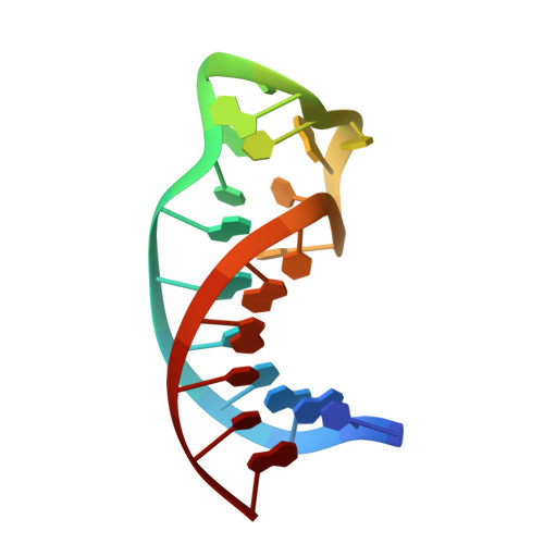
    1DB6

  • PubMed Abstract: 

    The structure of a DNA aptamer, which was selected for specific binding to arginine, was determined using NMR spectroscopy. The sequence forms a hairpin loop, with residues important for binding occurring in the loop region. Binding of argininamide induces formation of one Watson-Crick and two non-Watson-Crick base pairs, which facilitate generation of a binding pocket. The specificity for arginine seems to arise from contacts between the guanidino end of the arginine and phosphates, with atoms positioned by the shape of the pocket. Complex binding kinetics are observed suggesting that there is a slow interconversion of two forms of the DNA, which have different binding affinities. These data provide information on the process of adaptive recognition of a ligand by an aptamer.


  • Organizational Affiliation

    Department of Chemistry, University of California, Berkeley, California 94720-1460, USA.


Macromolecules

Find similar nucleic acids by:  Sequence   |   3D Structure  

Entity ID: 1
MoleculeChains LengthOrganismImage
DNA22N/A
Sequence Annotations
Expand
  • Reference Sequence
Small Molecules
Ligands 1 Unique
IDChains Name / Formula / InChI Key2D Diagram3D Interactions
AAR
Query on AAR

Download Ideal Coordinates CCD File 
B [auth A]ARGININEAMIDE
C6 H16 N5 O
ULEBESPCVWBNIF-BYPYZUCNSA-O
Binding Affinity Annotations 
IDSourceBinding Affinity
AAR PDBBind:  1DB6 Kd: 1.00e+5 (nM) from 1 assay(s)
Experimental Data & Validation

Experimental Data

  • Method: SOLUTION NMR
  • Conformers Calculated: 30 
  • Conformers Submitted: 10 
  • Selection Criteria: structures with acceptable covalent geometry,structures with favorable non- bond energy,structures with the least restraint violations,structures with the lowest energy 

Structure Validation

View Full Validation Report


Entry History 

Deposition Data

Revision History  (Full details and data files)

  • Version 1.0: 2000-02-03
    Type: Initial release
  • Version 1.1: 2008-04-27
    Changes: Version format compliance
  • Version 1.2: 2011-07-13
    Changes: Version format compliance
  • Version 1.3: 2022-02-16
    Changes: Data collection, Database references, Derived calculations