1CYO

BOVINE CYTOCHROME B(5)


Experimental Data Snapshot

  • Method: X-RAY DIFFRACTION
  • Resolution: 1.50 Å
  • R-Value Work: 0.160 
  • R-Value Observed: 0.160 

wwPDB Validation   3D Report Full Report


Ligand Structure Quality Assessment 


This is version 1.4 of the entry. See complete history


Literature

Refinement and structural analysis of bovine cytochrome b5 at 1.5 A resolution.

Durley, R.C.Mathews, F.S.

(1996) Acta Crystallogr D Biol Crystallogr 52: 65-76

  • DOI: https://doi.org/10.1107/S0907444995007827
  • Primary Citation of Related Structures:  
    1CYO

  • PubMed Abstract: 

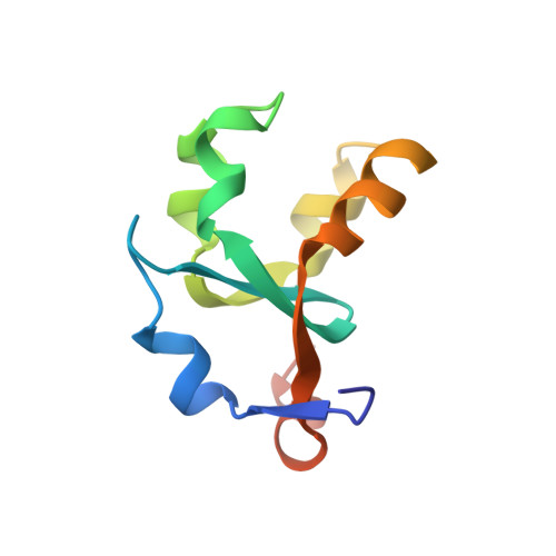
    The structure of bovine liver cytochrome b(5), a soluble 93-residue proteolytic fragment of a 16 kDa membrane-bound hemoprotein, initially solved at 2.0 A resolution, has been refined at 1.5 A using data collected on a diffractometer. Refinement to 2.0 A resolution used the Hendrickson-Konnert procedure PROLSQ and was then extended to 1.5 A resolution using the program PROFFT. Only residues 3-87 could be identified in the model and these residues together with 93 water molecules gave an agreement factor of R = 0.161 for data in the resolution range 1.5-5 A. The structure was finally refined using the program X-PLOR, which enabled alternate conformers to be modelled for several surface side chains. Residues 1 and 2 at the amino terminus of the protein and residue 88 near the carboxyl terminus could be identified from these electron-density maps. However the remaining disordered carboxy-terminal residues could not successfully be included in the model. A total of 117 solvent molecules were included in the final refinement to give R = 0.164 for the data between 1.5 and 10 A.


  • Organizational Affiliation

    Department of Biochemistry and Molecular Biology, Washington University School of Medicine, St. Louis, MO 63110, USA.


Macromolecules
Find similar proteins by:  (by identity cutoff)  |  3D Structure
Entity ID: 1
MoleculeChains Sequence LengthOrganismDetailsImage
CYTOCHROME B593Bos taurusMutation(s): 0 
UniProt
Find proteins for P00171 (Bos taurus)
Explore P00171 
Go to UniProtKB:  P00171
Entity Groups  
Sequence Clusters30% Identity50% Identity70% Identity90% Identity95% Identity100% Identity
UniProt GroupP00171
Sequence Annotations
Expand
  • Reference Sequence
Small Molecules
Ligands 1 Unique
IDChains Name / Formula / InChI Key2D Diagram3D Interactions
HEM
Query on HEM

Download Ideal Coordinates CCD File 
B [auth A]PROTOPORPHYRIN IX CONTAINING FE
C34 H32 Fe N4 O4
KABFMIBPWCXCRK-RGGAHWMASA-L
Experimental Data & Validation

Experimental Data

  • Method: X-RAY DIFFRACTION
  • Resolution: 1.50 Å
  • R-Value Work: 0.160 
  • R-Value Observed: 0.160 
  • Space Group: P 21 21 21
Unit Cell:
Length ( Å )Angle ( ˚ )
a = 64.54α = 90
b = 46.04β = 90
c = 29.91γ = 90
Software Package:
Software NamePurpose
X-PLORmodel building
X-PLORrefinement
X-PLORphasing

Structure Validation

View Full Validation Report


Ligand Structure Quality Assessment 


Entry History 

Deposition Data

Revision History  (Full details and data files)

  • Version 1.0: 1994-11-30
    Type: Initial release
  • Version 1.1: 2008-03-24
    Changes: Version format compliance
  • Version 1.2: 2011-07-13
    Changes: Version format compliance
  • Version 1.3: 2017-11-29
    Changes: Derived calculations, Other
  • Version 1.4: 2024-02-07
    Changes: Data collection, Database references, Derived calculations