1CR2

CRYSTAL STRUCTURE OF THE HELICASE DOMAIN OF THE GENE 4 PROTEIN OF BACTERIOPHAGE T7: COMPLEX WITH DATP


Experimental Data Snapshot

  • Method: X-RAY DIFFRACTION
  • Resolution: 2.30 Å
  • R-Value Free: 0.296 
  • R-Value Work: 0.245 
  • R-Value Observed: 0.250 

wwPDB Validation   3D Report Full Report


Ligand Structure Quality Assessment 


This is version 1.4 of the entry. See complete history


Literature

Crystal structure of the helicase domain from the replicative helicase-primase of bacteriophage T7.

Sawaya, M.R.Guo, S.Tabor, S.Richardson, C.C.Ellenberger, T.

(1999) Cell 99: 167-177

  • DOI: https://doi.org/10.1016/s0092-8674(00)81648-7
  • Primary Citation of Related Structures:  
    1CR0, 1CR1, 1CR2, 1CR4

  • PubMed Abstract: 

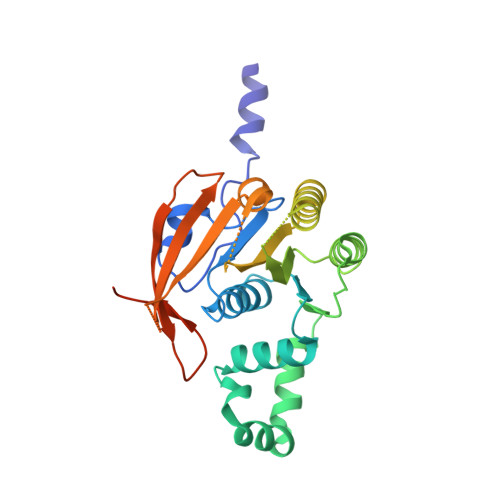
    Helicases that unwind DNA at the replication fork are ring-shaped oligomeric enzymes that move along one strand of a DNA duplex and catalyze the displacement of the complementary strand in a reaction that is coupled to nucleotide hydrolysis. The helicase domain of the replicative helicase-primase protein from bacteriophage T7 crystallized as a helical filament that resembles the Escherichia coli RecA protein, an ATP-dependent DNA strand exchange factor. When viewed in projection along the helical axis of the crystals, six protomers of the T7 helicase domain resemble the hexameric rings seen in electron microscopic images of the intact T7 helicase-primase. Nucleotides bind at the interface between pairs of adjacent subunits where an arginine is near the gamma-phosphate of the nucleotide in trans. The bound nucleotide stabilizes the folded conformation of a DNA-binding motif located near the center of the ring. These and other observations suggest how conformational changes are coupled to DNA unwinding activity.


  • Organizational Affiliation

    Department of Biological Chemistry and Molecular Pharmacology, Harvard Medical School, Boston, Massachusetts 02115, USA.


Macromolecules
Find similar proteins by:  (by identity cutoff)  |  3D Structure
Entity ID: 1
MoleculeChains Sequence LengthOrganismDetailsImage
DNA PRIMASE/HELICASE296Escherichia phage T7Mutation(s): 1 
EC: 2.7.7
UniProt
Find proteins for P03692 (Escherichia phage T7)
Explore P03692 
Go to UniProtKB:  P03692
Entity Groups  
Sequence Clusters30% Identity50% Identity70% Identity90% Identity95% Identity100% Identity
UniProt GroupP03692
Sequence Annotations
Expand
  • Reference Sequence
Experimental Data & Validation

Experimental Data

  • Method: X-RAY DIFFRACTION
  • Resolution: 2.30 Å
  • R-Value Free: 0.296 
  • R-Value Work: 0.245 
  • R-Value Observed: 0.250 
  • Space Group: P 61
Unit Cell:
Length ( Å )Angle ( ˚ )
a = 80.172α = 90
b = 80.172β = 90
c = 85.709γ = 120
Software Package:
Software NamePurpose
X-PLORmodel building
X-PLORrefinement
DENZOdata reduction
SCALEPACKdata scaling
X-PLORphasing

Structure Validation

View Full Validation Report


Ligand Structure Quality Assessment 


Entry History 

Deposition Data

Revision History  (Full details and data files)

  • Version 1.0: 1999-11-10
    Type: Initial release
  • Version 1.1: 2008-04-27
    Changes: Version format compliance
  • Version 1.2: 2011-07-13
    Changes: Version format compliance
  • Version 1.3: 2021-11-03
    Changes: Database references, Derived calculations
  • Version 1.4: 2024-02-07
    Changes: Data collection