1CAH

STRUCTURE OF COBALT CARBONIC ANHYDRASE COMPLEXED WITH BICARBONATE


Experimental Data Snapshot

  • Method: X-RAY DIFFRACTION
  • Resolution: 1.88 Å
  • R-Value Observed: 0.160 

wwPDB Validation   3D Report Full Report


This is version 1.4 of the entry. See complete history


Literature

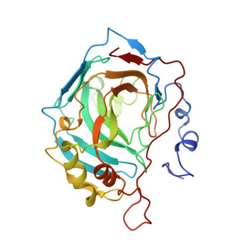
Structure of cobalt carbonic anhydrase complexed with bicarbonate.

Hakansson, K.Wehnert, A.

(1992) J Mol Biol 228: 1212-1218

  • DOI: https://doi.org/10.1016/0022-2836(92)90327-g
  • Primary Citation of Related Structures:  
    1CAH

  • PubMed Abstract: 

    The three-dimensional structure of a complex between catalytically active cobalt(II) substituted human carbonic anhydrase II and its substrate bicarbonate was determined by X-ray crystallography (1.9 A). One water molecule and two bicarbonate oxygen atoms are found at distances between 2.3 and 2.5 A from the cobalt ion in addition to the three histidyl ligands contributed by the peptide chain. The tetrahedral geometry around the metal ion in the native enzyme with a single water molecule 2.0 A from the metal is therefore lost. The geometry is difficult to classify but might best be described as distorted octahedral. The structure is suggested to represent a water-bicarbonate exchange state relevant also for native carbonic anhydrase, where the two unprotonized oxygen atoms of the substrate are bound in a carboxylate binding site and the hydroxyl group is free to move closer to the metal thereby replacing the metal-bound water molecule. A reaction mechanism based on crystallographically determined enzyme-ligand complexes is represented.


  • Organizational Affiliation

    Molecular Biophysics, Chemical Center, University of Lund, Sweden.


Macromolecules
Find similar proteins by:  (by identity cutoff)  |  3D Structure
Entity ID: 1
MoleculeChains Sequence LengthOrganismDetailsImage
CARBONIC ANHYDRASE II259Homo sapiensMutation(s): 0 
EC: 4.2.1.1
UniProt & NIH Common Fund Data Resources
Find proteins for P00918 (Homo sapiens)
Explore P00918 
Go to UniProtKB:  P00918
PHAROS:  P00918
GTEx:  ENSG00000104267 
Entity Groups  
Sequence Clusters30% Identity50% Identity70% Identity90% Identity95% Identity100% Identity
UniProt GroupP00918
Sequence Annotations
Expand
  • Reference Sequence
Small Molecules
Binding Affinity Annotations 
IDSourceBinding Affinity
BCT BindingDB:  1CAH Ki: min: 7.30e+7, max: 8.50e+7 (nM) from 2 assay(s)
Experimental Data & Validation

Experimental Data

  • Method: X-RAY DIFFRACTION
  • Resolution: 1.88 Å
  • R-Value Observed: 0.160 
  • Space Group: P 1 21 1
Unit Cell:
Length ( Å )Angle ( ˚ )
a = 42.7α = 90
b = 41.7β = 104.6
c = 73γ = 90
Software Package:
Software NamePurpose
PROFFTrefinement

Structure Validation

View Full Validation Report


Entry History 

Deposition Data

Revision History  (Full details and data files)

  • Version 1.0: 1993-10-31
    Type: Initial release
  • Version 1.1: 2008-03-03
    Changes: Version format compliance
  • Version 1.2: 2011-07-13
    Changes: Version format compliance
  • Version 1.3: 2017-11-29
    Changes: Derived calculations, Other
  • Version 1.4: 2024-02-07
    Changes: Data collection, Database references, Derived calculations