1BWU

MANNOSE-SPECIFIC AGGLUTININ (LECTIN) FROM GARLIC (ALLIUM SATIVUM) BULBS COMPLEXED WITH ALPHA-D-MANNOSE


Experimental Data Snapshot

  • Method: X-RAY DIFFRACTION
  • Resolution: 2.80 Å
  • R-Value Free: 0.278 
  • R-Value Work: 0.226 
  • R-Value Observed: 0.226 

wwPDB Validation   3D Report Full Report


Ligand Structure Quality Assessment 


This is version 1.5 of the entry. See complete history


Literature

Crystal structure of a dimeric mannose-specific agglutinin from garlic: quaternary association and carbohydrate specificity.

Chandra, N.R.Ramachandraiah, G.Bachhawat, K.Dam, T.K.Surolia, A.Vijayan, M.

(1999) J Mol Biol 285: 1157-1168

  • DOI: https://doi.org/10.1006/jmbi.1998.2353
  • Primary Citation of Related Structures:  
    1BWU

  • PubMed Abstract: 

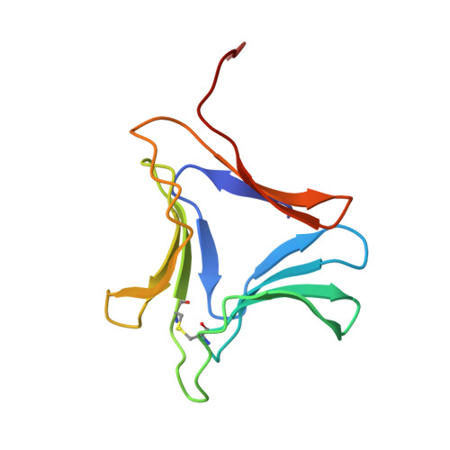
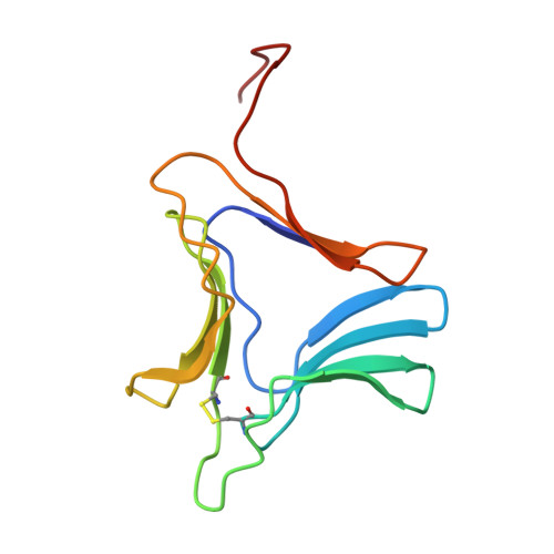
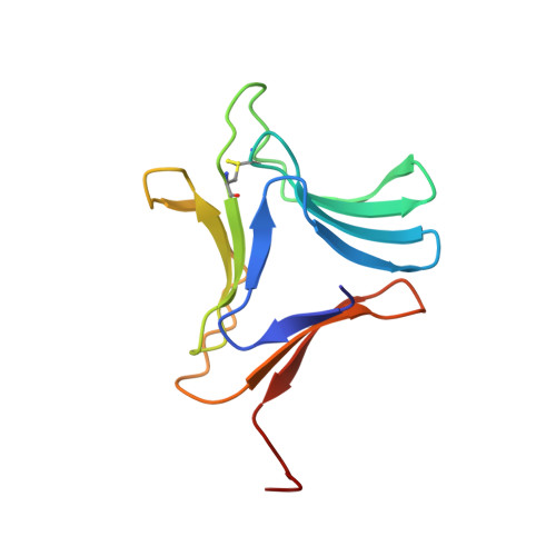
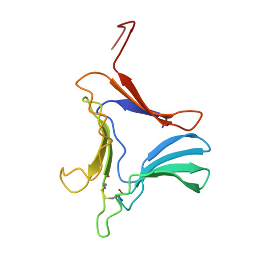
    A mannose-specific agglutinin, isolated from garlic bulbs, has been crystallized in the presence of a large excess of alpha-d-mannose, in space group C2 and cell dimensions, a=203.24, b=43.78, c=79.27 A, beta=112.4 degrees, with two dimers in the asymmetric unit. X-ray diffraction data were collected up to a nominal resolution of 2.4 A and the structure was solved by molecular replacement. The structure, refined to an R-factor of 22.6 % and an Rfree of 27.8 % reveals a beta-prism II fold, similar to that in the snowdrop lectin, comprising three antiparallel four-stranded beta-sheets arranged as a 12-stranded beta-barrel, with an approximate internal 3-fold symmetry. This agglutinin is, however, a dimer unlike snowdrop lectin which exists as a tetramer, despite a high degree of sequence similarity between them. A comparison of the two structures reveals a few substitutions in the garlic lectin which stabilise it into a dimer and prevent tetramer formation. Three mannose molecules have been identified on each subunit. In addition, electron density is observed for another possible mannose molecule per dimer resulting in a total of seven mannose molecules in each dimer. Although the mannose binding sites and the overall structure are similar in the subunits of snowdrop and garlic lectin, their specificities to glycoproteins such as GP120 vary considerably. These differences appear, in part, to be a direct consequence of the differences in oligomerisation, implying that variation in quaternary association may be a mode of achieving oligosaccharide specificity in bulb lectins.


  • Organizational Affiliation

    Molecular Biophysics Unit, Indian Institute of Science, Bangalore, 560 012, India.


Macromolecules
Find similar proteins by:  (by identity cutoff)  |  3D Structure
Entity ID: 1
MoleculeChains Sequence LengthOrganismDetailsImage
PROTEIN (AGGLUTININ)106Allium sativumMutation(s): 0 
UniProt
Find proteins for Q38789 (Allium sativum)
Explore Q38789 
Go to UniProtKB:  Q38789
Entity Groups  
Sequence Clusters30% Identity50% Identity70% Identity90% Identity95% Identity100% Identity
UniProt GroupQ38789
Sequence Annotations
Expand
  • Reference Sequence
Find similar proteins by:  (by identity cutoff)  |  3D Structure
Entity ID: 2
MoleculeChains Sequence LengthOrganismDetailsImage
PROTEIN (AGGLUTININ)B [auth D]109Allium sativumMutation(s): 0 
UniProt
Find proteins for Q38784 (Allium sativum)
Explore Q38784 
Go to UniProtKB:  Q38784
Entity Groups  
Sequence Clusters30% Identity50% Identity70% Identity90% Identity95% Identity100% Identity
UniProt GroupQ38784
Sequence Annotations
Expand
  • Reference Sequence
Find similar proteins by:  (by identity cutoff)  |  3D Structure
Entity ID: 3
MoleculeChains Sequence LengthOrganismDetailsImage
PROTEIN (AGGLUTININ)C [auth P]106Allium sativumMutation(s): 0 
UniProt
Find proteins for Q38789 (Allium sativum)
Explore Q38789 
Go to UniProtKB:  Q38789
Entity Groups  
Sequence Clusters30% Identity50% Identity70% Identity90% Identity95% Identity100% Identity
UniProt GroupQ38789
Sequence Annotations
Expand
  • Reference Sequence
Find similar proteins by:  (by identity cutoff)  |  3D Structure
Entity ID: 4
MoleculeChains Sequence LengthOrganismDetailsImage
PROTEIN (AGGLUTININ)D [auth Q]109Allium sativumMutation(s): 0 
UniProt
Find proteins for Q38784 (Allium sativum)
Explore Q38784 
Go to UniProtKB:  Q38784
Entity Groups  
Sequence Clusters30% Identity50% Identity70% Identity90% Identity95% Identity100% Identity
UniProt GroupQ38784
Sequence Annotations
Expand
  • Reference Sequence
Small Molecules
Ligands 1 Unique
IDChains Name / Formula / InChI Key2D Diagram3D Interactions
MAN
Query on MAN

Download Ideal Coordinates CCD File 
E [auth A]
F [auth A]
G [auth A]
H [auth D]
I [auth D]
E [auth A],
F [auth A],
G [auth A],
H [auth D],
I [auth D],
J [auth D],
K [auth D],
L [auth P],
M [auth P],
N [auth P],
O [auth Q],
P [auth Q],
Q,
R [auth Q]
alpha-D-mannopyranose
C6 H12 O6
WQZGKKKJIJFFOK-PQMKYFCFSA-N
Experimental Data & Validation

Experimental Data

  • Method: X-RAY DIFFRACTION
  • Resolution: 2.80 Å
  • R-Value Free: 0.278 
  • R-Value Work: 0.226 
  • R-Value Observed: 0.226 
  • Space Group: C 1 2 1
Unit Cell:
Length ( Å )Angle ( ˚ )
a = 203.243α = 90
b = 43.78β = 112.37
c = 79.268γ = 90
Software Package:
Software NamePurpose
XDSdata scaling
AUTOMARdata reduction
AMoREphasing
X-PLORrefinement
XDSdata reduction

Structure Validation

View Full Validation Report


Ligand Structure Quality Assessment 


Entry History 

Deposition Data

Revision History  (Full details and data files)

  • Version 1.0: 1999-01-20
    Type: Initial release
  • Version 1.1: 2008-04-27
    Changes: Version format compliance
  • Version 1.2: 2011-07-13
    Changes: Derived calculations, Version format compliance
  • Version 1.3: 2017-10-04
    Changes: Refinement description
  • Version 1.4: 2020-07-29
    Type: Remediation
    Reason: Carbohydrate remediation
    Changes: Advisory, Data collection, Database references, Derived calculations, Structure summary
  • Version 1.5: 2023-08-09
    Changes: Data collection, Database references, Refinement description, Structure summary