1BVN

PIG PANCREATIC ALPHA-AMYLASE IN COMPLEX WITH THE PROTEINACEOUS INHIBITOR TENDAMISTAT


Experimental Data Snapshot

  • Method: X-RAY DIFFRACTION
  • Resolution: 2.50 Å
  • R-Value Free: 0.260 
  • R-Value Work: 0.166 
  • R-Value Observed: 0.166 

wwPDB Validation   3D Report Full Report


This is version 1.2 of the entry. See complete history


Literature

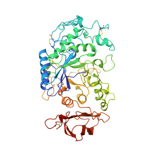
The crystal structure of porcine pancreatic alpha-amylase in complex with the microbial inhibitor Tendamistat.

Wiegand, G.Epp, O.Huber, R.

(1995) J Mol Biol 247: 99-110

  • DOI: https://doi.org/10.1006/jmbi.1994.0125
  • Primary Citation of Related Structures:  
    1BVN

  • PubMed Abstract: 

    The crystal structure of the complex formed between the 498 amino acid residue porcine pancreatic alpha-amylase (PPA) and the 74 amino acid residue inhibitor Tendamistat secreted from Streptomyces tendae, has been determined by multiple isomorphous replacement in a crystal of space group P6(5)22 (a = b = 77.7 A, c = 359.5 A). The model has been refined to an R-factor of 0.194 by Powell minimization applying strong energy constraints based on 17,964 independent reflections in the 7 to 2.5 A resolution range, and obeys standard geometry within 0.011 A in bond lengths and 1.78 degrees in bond angles. The final model consists of all 496 amino acid residues of PPA, 71 amino acid residues of Tendamistat (without the three N-terminal residues), one calcium ion, one chloride ion and 167 water molecules. PPA exhibits the same topological fold in the complex as the uncomplexed PPA recently published by others. About 30% of the water-accessible surface of Tendamistat is in contact with PPA. Four segments of the polypeptide chain, with a total of 15 amino acid residues, are involved in the binding. One segment containing the staggered side-chains of the triplet Trp18, Arg19, Tyr20, typical for this class of inhibitors, binds into the catalytic site. The other segments fill out the groove in the PPA molecule, which also binds the carbohydrate inhibitor acarbose and is assumed to be the substrate-binding region. This extended interaction between Tendamistat and alpha-amylase explains the very high inhibition constant of about 9 x 10(-12) M.


  • Organizational Affiliation

    Max-Planck-Institut für Biochemie, Martinsried, F.R.G.


Macromolecules
Find similar proteins by:  (by identity cutoff)  |  3D Structure
Entity ID: 1
MoleculeChains Sequence LengthOrganismDetailsImage
PROTEIN (ALPHA-AMYLASE)A [auth P]496Sus scrofaMutation(s): 0 
EC: 3.2.1.1
UniProt
Find proteins for P00690 (Sus scrofa)
Explore P00690 
Go to UniProtKB:  P00690
Entity Groups  
Sequence Clusters30% Identity50% Identity70% Identity90% Identity95% Identity100% Identity
UniProt GroupP00690
Sequence Annotations
Expand
  • Reference Sequence
Find similar proteins by:  (by identity cutoff)  |  3D Structure
Entity ID: 2
MoleculeChains Sequence LengthOrganismDetailsImage
PROTEIN (TENDAMISTAT)B [auth T]74Streptomyces tendaeMutation(s): 0 
UniProt
Find proteins for P01092 (Streptomyces tendae)
Explore P01092 
Go to UniProtKB:  P01092
Entity Groups  
Sequence Clusters30% Identity50% Identity70% Identity90% Identity95% Identity100% Identity
UniProt GroupP01092
Sequence Annotations
Expand
  • Reference Sequence
Small Molecules
Ligands 2 Unique
IDChains Name / Formula / InChI Key2D Diagram3D Interactions
CA
Query on CA

Download Ideal Coordinates CCD File 
C [auth P]CALCIUM ION
Ca
BHPQYMZQTOCNFJ-UHFFFAOYSA-N
CL
Query on CL

Download Ideal Coordinates CCD File 
D [auth P]CHLORIDE ION
Cl
VEXZGXHMUGYJMC-UHFFFAOYSA-M
Experimental Data & Validation

Experimental Data

  • Method: X-RAY DIFFRACTION
  • Resolution: 2.50 Å
  • R-Value Free: 0.260 
  • R-Value Work: 0.166 
  • R-Value Observed: 0.166 
  • Space Group: P 65 2 2
Unit Cell:
Length ( Å )Angle ( ˚ )
a = 77.7α = 90
b = 77.7β = 90
c = 359.5γ = 120
Software Package:
Software NamePurpose
FILMEdata collection
PROTEINdata reduction
PROTEINmodel building
X-PLORrefinement
FILMEdata reduction
PROTEINdata scaling
PROTEINphasing

Structure Validation

View Full Validation Report


Entry History 

Deposition Data

Revision History  (Full details and data files)

  • Version 1.0: 1998-09-23
    Type: Initial release
  • Version 1.1: 2007-10-16
    Changes: Version format compliance
  • Version 1.2: 2011-07-13
    Changes: Version format compliance