1BM9

REPLICATION TERMINATOR PROTEIN FROM BACILLUS SUBTILIS


Experimental Data Snapshot

  • Method: X-RAY DIFFRACTION
  • Resolution: 2.00 Å
  • R-Value Free: 0.281 
  • R-Value Work: 0.206 
  • R-Value Observed: 0.206 

wwPDB Validation   3D Report Full Report


This is version 1.3 of the entry. See complete history


Literature

Crystal structure of the replication terminator protein from B. subtilis at 2.6 A.

Bussiere, D.E.Bastia, D.White, S.W.

(1995) Cell 80: 651-660

  • DOI: https://doi.org/10.1016/0092-8674(95)90519-7
  • Primary Citation of Related Structures:  
    1BM9

  • PubMed Abstract: 

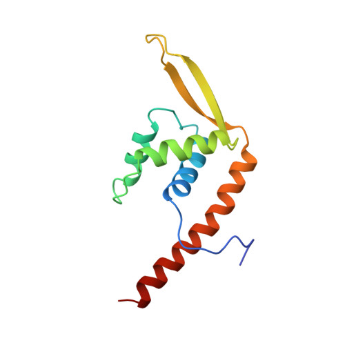
    The crystal structure of the replication terminator protein (RTP) of B. subtilis has been determined at 2.6 A resolution. As previously suggested by both biochemical and biophysical studies, the molecule exists as a symmetric dimer and is in the alpha + beta protein-folding class. The protein has several uncommon features, including an antiparallel coiled-coil, which serves as the dimerization domain, and both an alpha-helix and a beta-ribbon suitably positioned to interact with the major and minor grooves of B-DNA. A site has been identified on the surface of RTP that is biochemically and positionally suitable for interaction with the replication-specific helicase. Other features of the structure are consistent with the polar contrahelicase mechanism of the protein. A model of the interaction between RTP and its cognate DNA is presented.


  • Organizational Affiliation

    Department of Microbiology, Duke University Medical Center, Durham, North Carolina 27710.


Macromolecules
Find similar proteins by:  (by identity cutoff)  |  3D Structure
Entity ID: 1
MoleculeChains Sequence LengthOrganismDetailsImage
REPLICATION TERMINATOR PROTEIN
A, B
122Bacillus subtilisMutation(s): 0 
Gene Names: RTP
UniProt
Find proteins for P0CI76 (Bacillus subtilis (strain 168))
Explore P0CI76 
Go to UniProtKB:  P0CI76
Entity Groups  
Sequence Clusters30% Identity50% Identity70% Identity90% Identity95% Identity100% Identity
UniProt GroupP0CI76
Sequence Annotations
Expand
  • Reference Sequence
Experimental Data & Validation

Experimental Data

  • Method: X-RAY DIFFRACTION
  • Resolution: 2.00 Å
  • R-Value Free: 0.281 
  • R-Value Work: 0.206 
  • R-Value Observed: 0.206 
  • Space Group: C 1 2 1
Unit Cell:
Length ( Å )Angle ( ˚ )
a = 77.191α = 90
b = 52.686β = 90.52
c = 70.583γ = 90
Software Package:
Software NamePurpose
PHASESphasing
CNSrefinement
R-AXISdata reduction
R-AXISdata scaling

Structure Validation

View Full Validation Report


Entry History 

Deposition Data

Revision History  (Full details and data files)

  • Version 1.0: 1999-01-06
    Type: Initial release
  • Version 1.1: 2008-03-27
    Changes: Version format compliance
  • Version 1.2: 2011-07-13
    Changes: Derived calculations, Version format compliance
  • Version 1.3: 2024-02-07
    Changes: Data collection, Database references, Structure summary