1AVY

FIBRITIN DELETION MUTANT M (BACTERIOPHAGE T4)


Experimental Data Snapshot

  • Method: X-RAY DIFFRACTION
  • Resolution: 1.85 Å
  • R-Value Free: 0.253 
  • R-Value Work: 0.220 
  • R-Value Observed: 0.220 

wwPDB Validation   3D Report Full Report


This is version 1.5 of the entry. See complete history


Literature

Structure of bacteriophage T4 fibritin M: a troublesome packing arrangement.

Strelkov, S.V.Tao, Y.Shneider, M.M.Mesyanzhinov, V.V.Rossmann, M.G.

(1998) Acta Crystallogr D Biol Crystallogr 54: 805-816

  • DOI: https://doi.org/10.1107/s0907444997018878
  • Primary Citation of Related Structures:  
    1AVY

  • PubMed Abstract: 

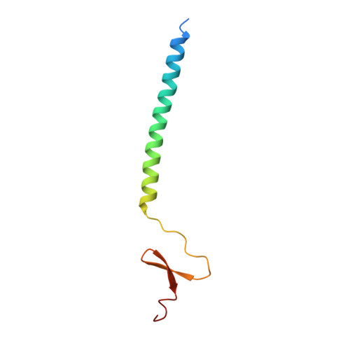
    Fibritin, a 52 kDa product of bacteriophage T4 gene wac, forms 530 A long fibers, named whiskers, that attach to the phage neck and perform a helper function during phage assembly. Fibritin is a homotrimer, with its predominant central domain consisting of 12 consecutive alpha-helical coiled-coil segments linked together by loops. The central domain is flanked by small globular domains at both ends. Fibritin M is a genetically engineered fragment of the wild type and contains 74 amino-acid residues corresponding to the last coiled-coil segment and the complete carboxy-terminal domain. The crystals of fibritin M belong to the rare space group P3 with three crystallographically independent trimers in the unit cell. The structure has been established at 1.85 A resolution by combining molecular and isomorphous replacement techniques. One of the two heavy-atom derivatives used was gaseous xenon. A substantial fraction of residues in each independent trimer is disordered to various extents in proportion to the lack of restraints on the molecules provided by the lattice contacts. Accurate modeling of the solvent present in the crystals was crucial for achieving good agreement with experimental data.


  • Organizational Affiliation

    Department of Biological Sciences, Purdue University, West Lafayette, IN 47907-1392, USA.


Macromolecules
Find similar proteins by:  (by identity cutoff)  |  3D Structure
Entity ID: 1
MoleculeChains Sequence LengthOrganismDetailsImage
FIBRITIN
A, B, C
74Tequatrovirus T4Mutation(s): 4 
Gene Names: WAC
UniProt
Find proteins for P10104 (Enterobacteria phage T4)
Explore P10104 
Go to UniProtKB:  P10104
Entity Groups  
Sequence Clusters30% Identity50% Identity70% Identity90% Identity95% Identity100% Identity
UniProt GroupP10104
Sequence Annotations
Expand
  • Reference Sequence
Experimental Data & Validation

Experimental Data

  • Method: X-RAY DIFFRACTION
  • Resolution: 1.85 Å
  • R-Value Free: 0.253 
  • R-Value Work: 0.220 
  • R-Value Observed: 0.220 
  • Space Group: P 3
Unit Cell:
Length ( Å )Angle ( ˚ )
a = 43.68α = 90
b = 43.68β = 90
c = 90.61γ = 120
Software Package:
Software NamePurpose
X-PLORmodel building
X-PLORrefinement
DENZOdata reduction
SCALEPACKdata scaling
X-PLORphasing

Structure Validation

View Full Validation Report


Entry History 

Deposition Data

Revision History  (Full details and data files)

  • Version 1.0: 1997-12-03
    Type: Initial release
  • Version 1.1: 2008-03-24
    Changes: Version format compliance
  • Version 1.2: 2011-07-13
    Changes: Version format compliance
  • Version 1.3: 2011-11-16
    Changes: Atomic model
  • Version 1.4: 2021-11-03
    Changes: Database references, Other
  • Version 1.5: 2023-08-02
    Changes: Refinement description