Three-dimensional structure in solution of neurotoxin III from the sea anemone Anemonia sulcata.
Manoleras, N., Norton, R.S.(1994) Biochemistry 33: 11051-11061
- PubMed: 7727358
- DOI: https://doi.org/10.1021/bi00203a001
- Primary Citation of Related Structures:
1ANS - PubMed Abstract:
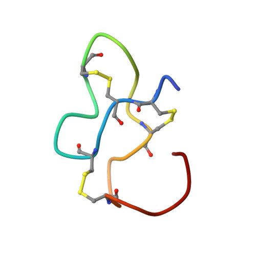
The three-dimensional structure in aqueous solution of the 27-residue polypeptide neurotoxin Anemonia sulcata toxin III (ATX III) has been determined from 1H NMR data. As ATX III self-associates in the millimolar concentration range, causing a marked concentration dependence for the chemical shifts of several residues [Norton, R. S., Cross, K., Braach-Maksvytis, V., & Wachter, E. (1993) Biochem. J. 293, 545-551], it was necessary to record NOESY spectra over a range of concentrations in order to eliminate any intermolecular interactions from the NOE restraint set. The pairings of the six half-cystine residues were also unknown and had to be determined (as 3-17, 4-11, and 6-22) from preliminary structure calculations performed using both upper bound distance restraints from NOESY data and a substantial number of lower bound restraints inferred from the absence of NOESY cross-peaks. Final structures were determined, using the program X-PLOR, from interproton distance restraints inferred from NOEs, backbone and side chain dihedral angle restraints from spin-spin coupling measurements, and a smaller number of lower bound restraints. Stereospecific assignments for 11 beta-methylene pairs were also included. The final set of 28 structures had an average pairwise RMS difference of 1.32 A over the backbone heavy atoms (N, C alpha, and C) and 2.18 A over all heavy atoms. For the well-defined region encompassing residues 3-22, the corresponding values were 0.62 and 1.28 A, respectively. ATX III adopts a compact structure containing four reverse turns (a distorted type I beta-turn at residues 6-9, a type I beta-turn at residues 8-11, and inverse gamma-turns at residues 12-14 and 15-17) and two other chain reversals, but no regular alpha-helix or beta-sheet. Several of the residues most affected by aggregation are located on the surface of the molecule, forming a hydrophobic patch which may constitute part of the sodium channel binding surface. Possible relationships between the structure of ATX III and those of other sea anemone toxins that interact with the same site on the voltage-gated sodium channel are considered.
Organizational Affiliation:
School of Biochemistry and Molecular Genetics, University of New South Wales, Kensington, Australia.