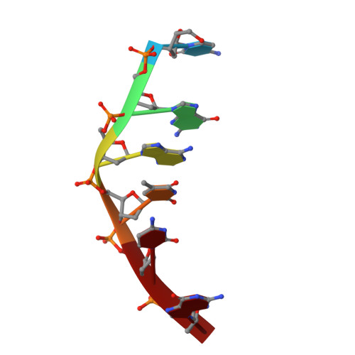
Structure of a DNA-bisdaunomycin complex.
Hu, G.G., Shui, X., Leng, F., Priebe, W., Chaires, J.B., Williams, L.D.(1997) Biochemistry 36: 5940-5946
- PubMed: 9166763
- DOI: https://doi.org/10.1021/bi9705218
- Primary Citation of Related Structures:
1AGL - PubMed Abstract:
The application of detailed structural data bases has now culminated in the successful design of a new generation of bisanthracyclines that form ultratight DNA complexes [Chaires, J. B., Leng, F., Przewloka, T., Fokt, I., Ling, Y. H., Perez-Soler, R., & Priebe, W. (1997) J. Med. Chem. 40, 261-266]. Daunomycin dimers were designed to bind to DNA in complexes resembling those of monomers intercalated at adjacent sites. The goal of the work described here was to determine, with X-ray crystallography, if a potent member of this newly designed and synthesized class of bisanthracyclines (WP631) binds as intended. WP631 is composed of two daunomycin molecules, linked N3' to N3' by a xylyl group. We have solved the 2.2 A X-ray crystal structure of a complex of WP631 bound to [d(CGATCG)]2. We demonstrate, on a detailed molecular level, that the WP631 design strategy is a success. The structures of WP631 and two daunomycin molecules bound to [d(CGATCG)]2 provide the unprecedented opportunity for detailed comparison of mono- and bis-intercalated complexes of the same chromophore, allowing us to distinguish effects of mono-intercalation from those of bis-intercalation. Differences are focused primarily in the centers of the complexes. DNA unwinding and other helical distortions propagate more efficiently to the center of the WP631 complex than to the center of the daunomycin complex.
Organizational Affiliation:
School of Chemistry & Biochemistry, Georgia Institute of Technology, Atlanta 30332-0400, USA.