1A4Y

RIBONUCLEASE INHIBITOR-ANGIOGENIN COMPLEX


Experimental Data Snapshot

  • Method: X-RAY DIFFRACTION
  • Resolution: 2.00 Å
  • R-Value Free: 0.286 
  • R-Value Work: 0.193 
  • R-Value Observed: 0.193 

wwPDB Validation   3D Report Full Report


This is version 1.3 of the entry. See complete history


Literature

Molecular recognition of human angiogenin by placental ribonuclease inhibitor--an X-ray crystallographic study at 2.0 A resolution.

Papageorgiou, A.C.Shapiro, R.Acharya, K.R.

(1997) EMBO J 16: 5162-5177

  • DOI: https://doi.org/10.1093/emboj/16.17.5162
  • Primary Citation of Related Structures:  
    1A4Y

  • PubMed Abstract: 

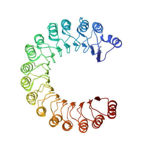
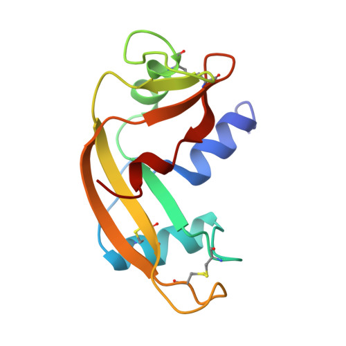
    Human placental RNase inhibitor (hRI), a leucine-rich repeat protein, binds the blood vessel-inducing protein human angiogenin (Ang) with extraordinary affinity (Ki <1 fM). Here we report a 2.0 A resolution crystal structure for the hRI-Ang complex that, together with extensive mutagenesis data from earlier studies, reveals the molecular features of this tight interaction. The hRI-Ang binding interface is large and encompasses 26 residues from hRI and 24 from Ang, recruited from multiple domains of both proteins. However, a substantial fraction of the energetically important contacts involve only a single region of each: the C-terminal segment 434-460 of hRI and the ribonucleolytic active centre of Ang, most notably the catalytic residue Lys40. Although the overall docking of Ang resembles that observed for RNase A in the crystal structure of its complex with the porcine RNase inhibitor, the vast majority of the interactions in the two complexes are distinctive, indicating that the broad specificity of the inhibitor for pancreatic RNase superfamily proteins is based largely on its capacity to recognize features unique to each of them. The implications of these findings for the development of small, hRI-based inhibitors of Ang for therapeutic use are discussed.


  • Organizational Affiliation

    Department of Biology and Biochemistry, University of Bath, Claverton Down, Bath BA2 7AY, UK.


Macromolecules
Find similar proteins by:  (by identity cutoff)  |  3D Structure
Entity ID: 1
MoleculeChains Sequence LengthOrganismDetailsImage
RIBONUCLEASE INHIBITORA,
C [auth D]
460Homo sapiensMutation(s): 0 
UniProt & NIH Common Fund Data Resources
Find proteins for P13489 (Homo sapiens)
Explore P13489 
Go to UniProtKB:  P13489
PHAROS:  P13489
GTEx:  ENSG00000023191 
Entity Groups  
Sequence Clusters30% Identity50% Identity70% Identity90% Identity95% Identity100% Identity
UniProt GroupP13489
Sequence Annotations
Expand
  • Reference Sequence
Find similar proteins by:  (by identity cutoff)  |  3D Structure
Entity ID: 2
MoleculeChains Sequence LengthOrganismDetailsImage
ANGIOGENINB,
D [auth E]
123Homo sapiensMutation(s): 0 
EC: 3.1.27
UniProt & NIH Common Fund Data Resources
Find proteins for P03950 (Homo sapiens)
Explore P03950 
Go to UniProtKB:  P03950
PHAROS:  P03950
GTEx:  ENSG00000214274 
Entity Groups  
Sequence Clusters30% Identity50% Identity70% Identity90% Identity95% Identity100% Identity
UniProt GroupP03950
Sequence Annotations
Expand
  • Reference Sequence
Experimental Data & Validation

Experimental Data

  • Method: X-RAY DIFFRACTION
  • Resolution: 2.00 Å
  • R-Value Free: 0.286 
  • R-Value Work: 0.193 
  • R-Value Observed: 0.193 
  • Space Group: P 1 21 1
Unit Cell:
Length ( Å )Angle ( ˚ )
a = 66.553α = 90
b = 105.605β = 107.09
c = 93.516γ = 90
Software Package:
Software NamePurpose
DENZOdata reduction
XDSdata scaling
SCALAdata scaling
X-PLORmodel building
X-PLORrefinement
XDSdata reduction
CCP4data scaling
X-PLORphasing

Structure Validation

View Full Validation Report


Entry History 

Deposition Data

Revision History  (Full details and data files)

  • Version 1.0: 1998-10-14
    Type: Initial release
  • Version 1.1: 2008-03-24
    Changes: Version format compliance
  • Version 1.2: 2011-07-13
    Changes: Version format compliance
  • Version 1.3: 2023-08-02
    Changes: Database references, Refinement description