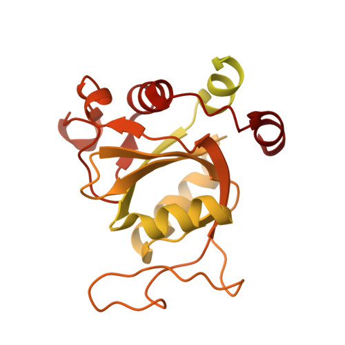
Atomistic and Thermodynamic Analysis of N6-Methyladenosine (m 6 A) Recognition by the Reader Domain of YTHDC1.
Li, Y., Bedi, R.K., Wiedmer, L., Sun, X., Huang, D., Caflisch, A.(2021) J Chem Theory Comput 17: 1240-1249
- PubMed: 33472367
- DOI: https://doi.org/10.1021/acs.jctc.0c01136
- Primary Citation of Related Structures:
6YNN, 6YNO, 6ZCM, 6ZCN, 6ZD3, 6ZD4, 6ZD5, 6ZD7, 6ZD8, 6ZD9, 6ZDA - PubMed Abstract:
N6-Methyladenosine (m 6 A) is the most frequent modification in eukaryotic messenger RNA (mRNA) and its cellular processing and functions are regulated by the reader proteins YTHDCs and YTHDFs. However, the mechanism of m 6 A recognition by the reader proteins is still elusive. Here, we investigate this recognition process by combining atomistic simulations, site-directed mutagenesis, and biophysical experiments using YTHDC1 as a model. We find that the N6 methyl group of m 6 A contributes to the binding through its specific interactions with an aromatic cage (formed by Trp377 and Trp428) and also by favoring the association-prone conformation of m 6 A-containing RNA in solution. The m 6 A binding site dynamically equilibrates between multiple metastable conformations with four residues being involved in the regulation of m 6 A binding (Trp428, Met438, Ser378, and Thr379). Trp428 switches between two conformational states to build and dismantle the aromatic cage. Interestingly, mutating Met438 and Ser378 to alanine does not alter m 6 A binding to the protein but significantly redistributes the binding enthalpy and entropy terms, i.e., enthalpy-entropy compensation. Such compensation is reasoned by different entropy-enthalpy transduction associated with both conformational changes of the wild-type and mutant proteins and the redistribution of water molecules. In contrast, the point mutant Thr379Val significantly changes the thermal stability and binding capability of YTHDC1 to its natural ligand. Additionally, thermodynamic analysis and free energy calculations shed light on the role of a structural water molecule that synergistically binds to YTHDC1 with m 6 A and acts as the hub of a hydrogen-bond network. Taken together, the experimental data and simulation results may accelerate the discovery of chemical probes, m 6 A-editing tools, and drug candidates against reader proteins.
Organizational Affiliation:
Department of Biochemistry, University of Zurich, Winterthurerstrasse 190, CH-8057 Zurich, Switzerland.


