6YCS

Human Transcription Cofactor PC4 DNA-binding domain in complex with full phosphorothioate 5-10-5 2'-O-methyl DNA gapmer antisense oligonucleotide.


Experimental Data Snapshot

  • Method: X-RAY DIFFRACTION
  • Resolution: 3.05 Å
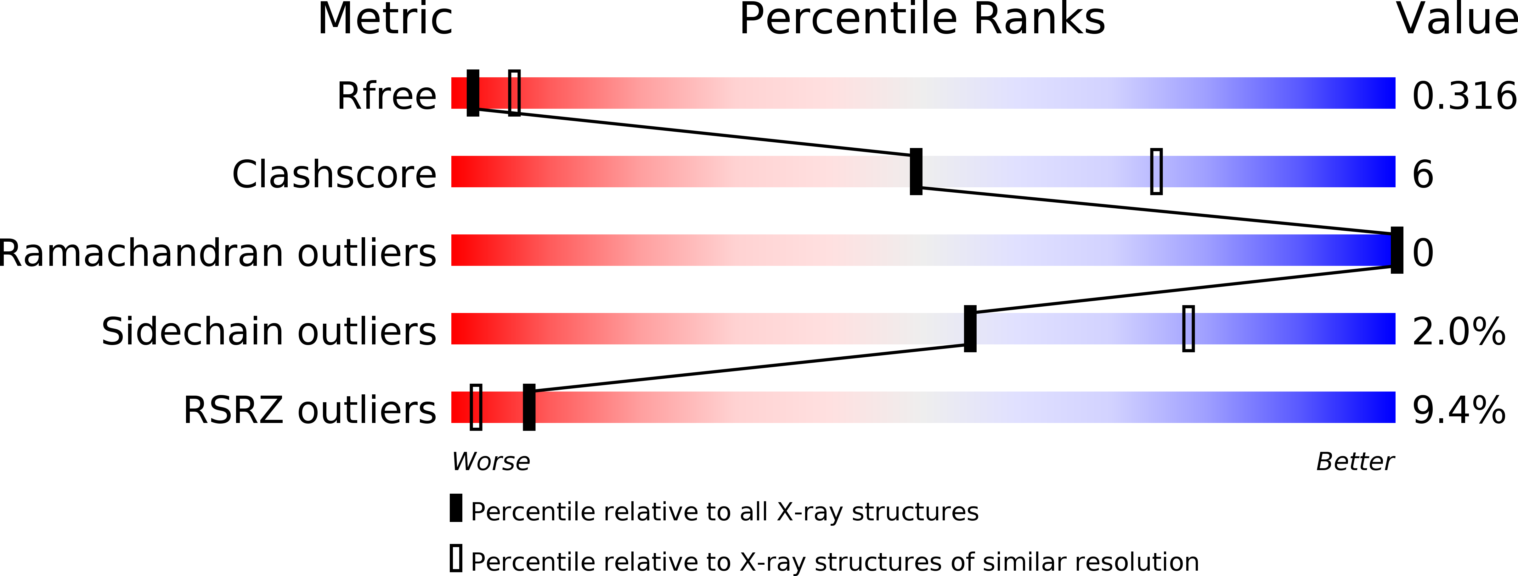
  • R-Value Free: 0.318 
  • R-Value Work: 0.238 
  • R-Value Observed: 0.241 

wwPDB Validation   3D Report Full Report


This is version 2.1 of the entry. See complete history


Literature

Origins of the Increased Affinity of Phosphorothioate-Modified Therapeutic Nucleic Acids for Proteins.

Hyjek-Skladanowska, M.Vickers, T.A.Napiorkowska, A.Anderson, B.A.Tanowitz, M.Crooke, S.T.Liang, X.H.Seth, P.P.Nowotny, M.

(2020) J Am Chem Soc 142: 7456-7468

  • DOI: https://doi.org/10.1021/jacs.9b13524
  • Primary Citation of Related Structures:  
    6YCS

  • PubMed Abstract: 

    The phosphorothioate backbone modification (PS) is one of the most widely used chemical modifications for enhancing the drug-like properties of nucleic acid-based drugs, including antisense oligonucleotides (ASOs). PS-modified nucleic acid therapeutics show improved metabolic stability from nuclease-mediated degradation and exhibit enhanced interactions with plasma, cell-surface, and intracellular proteins, which facilitates their tissue distribution and cellular uptake in animals. However, little is known about the structural basis of the interactions of PS nucleic acids with proteins. Here, we report a crystal structure of the DNA-binding domain of a model ASO-binding protein PC4, in complex with a full PS 2'-OMe DNA gapmer ASO. To our knowledge this is the first structure of a complex between a protein and fully PS nucleic acid. Each PC4 dimer comprises two DNA-binding interfaces. In the structure one interface binds the 5'-terminal 2'-OMe PS flank of the ASO, while the other interface binds the regular PS DNA central part in the opposite polarity. As a result, the ASO forms a hairpin-like structure. ASO binding also induces the formation of a dimer of dimers of PC4, which is stabilized by base pairing between homologous regions of the ASOs bound by each dimer of PC4. The protein interacts with the PS nucleic acid through a network of electrostatic and hydrophobic interactions, which provides insights into the origins for the enhanced affinity of PS for proteins. The importance of these contacts was further confirmed in a NanoBRET binding assay using a Nano luciferase tagged PC4 acting as the BRET donor, to a fluorescently conjugated ASO acting as the BRET acceptor. Overall, our results provide insights into the molecular forces that govern the interactions of PS ASOs with cellular proteins and provide a potential model for how these interactions can template protein-protein interactions causative of cellular toxicity.


  • Organizational Affiliation

    Structural Biology Center, International Institute of Molecular and Cell Biology, 4 Trojdena St., 02-109 Warsaw, Poland.


Macromolecules

Find similar proteins by:  (by identity cutoff)  |  3D Structure
Entity ID: 1
MoleculeChains Sequence LengthOrganismDetailsImage
PC4 protein
A, B, C, D
72Homo sapiensMutation(s): 0 
Gene Names: PC4SUB1hCG_1781938
UniProt & NIH Common Fund Data Resources
Find proteins for P53999 (Homo sapiens)
Explore P53999 
Go to UniProtKB:  P53999
PHAROS:  P53999
GTEx:  ENSG00000113387 
Entity Groups  
Sequence Clusters30% Identity50% Identity70% Identity90% Identity95% Identity100% Identity
UniProt GroupP53999
Sequence Annotations
Expand
  • Reference Sequence

Find similar nucleic acids by:  Sequence   |   3D Structure  

Entity ID: 2
MoleculeChains LengthOrganismImage
DNA (5'-D(P*(OKQ))-D(P*(OKT))-R(P*(RFJ))-D(*(OKQ)P*(OKT)P*(AS)P*(GS)P*(OKN)P*(OKN)P*(PST)P*(OKN)P*(PST)P*(GS)P*(GS)P*(AS)P*(OKT)P*(OKT))-3')17synthetic construct
Sequence Annotations
Expand
  • Reference Sequence

Find similar nucleic acids by:  Sequence   |   3D Structure  

Entity ID: 3
MoleculeChains LengthOrganismImage
DNA (5'-D(P*(OKQ))-D(P*(OKT))-R(P*(RFJ))-D(*(OKQ)P*(OKT)P*(AS)P*(GS)P*(OKN)P*(OKN)P*(PST)P*(OKN)P*(PST)P*(GS)P*(GS)P*(AS)P*(OKT)P*(OKT))-3')17synthetic construct
Sequence Annotations
Expand
  • Reference Sequence
Experimental Data & Validation

Experimental Data

  • Method: X-RAY DIFFRACTION
  • Resolution: 3.05 Å
  • R-Value Free: 0.318 
  • R-Value Work: 0.238 
  • R-Value Observed: 0.241 
  • Space Group: P 43 21 2
Unit Cell:
Length ( Å )Angle ( ˚ )
a = 102.143α = 90
b = 102.143β = 90
c = 83.813γ = 90
Software Package:
Software NamePurpose
PHENIXrefinement
XDSdata reduction
XDSdata scaling
PHASERphasing

Structure Validation

View Full Validation Report


Entry History 

Deposition Data

Revision History  (Full details and data files)

  • Version 1.0: 2020-04-29
    Type: Initial release
  • Version 1.1: 2020-05-06
    Changes: Structure summary
  • Version 2.0: 2020-11-11
    Changes: Advisory, Atomic model, Author supporting evidence, Data collection, Derived calculations, Non-polymer description, Polymer sequence, Source and taxonomy, Structure summary
  • Version 2.1: 2024-01-24
    Changes: Data collection, Database references, Refinement description