6Y40

14-3-3 Sigma in complex with phosphorylated PLN peptide


Experimental Data Snapshot

  • Method: X-RAY DIFFRACTION
  • Resolution: 1.75 Å
  • R-Value Free: 0.207 
  • R-Value Work: 0.168 
  • R-Value Observed: 0.170 

wwPDB Validation   3D Report Full Report


This is version 1.3 of the entry. See complete history


Literature

A new soaking procedure for X-ray crystallographic structural determination of protein-peptide complexes.

Ballone, A.Lau, R.A.Zweipfenning, F.P.A.Ottmann, C.

(2020) Acta Crystallogr F Struct Biol Commun 76: 501-507

  • DOI: https://doi.org/10.1107/S2053230X2001122X
  • Primary Citation of Related Structures:  
    6Y3M, 6Y3O, 6Y3R, 6Y3S, 6Y3V, 6Y40, 6Y44, 6Y8A, 6Y8B, 6Y8D, 6Y8E

  • PubMed Abstract: 

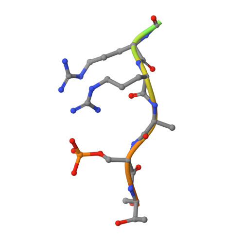
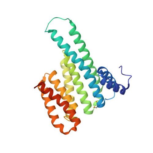
    Interactions between a protein and a peptide motif of its protein partner are prevalent in nature. Often, a protein also has multiple interaction partners. X-ray protein crystallography is commonly used to examine these interactions in terms of bond distances and angles as well as to describe hotspots within protein complexes. However, the crystallization process presents a significant bottleneck in structure determination since it often requires notably time-consuming screening procedures, which involve testing a broad range of crystallization conditions via a trial-and-error approach. This difficulty is also increased as each protein-peptide complex does not necessarily crystallize under the same conditions. Here, a new co-crystallization/peptide-soaking method is presented which circumvents the need to return to the initial lengthy crystal screening and optimization processes for each consequent new complex. The 14-3-3σ protein, which has multiple interacting partners with specific peptidic motifs, was used as a case study. It was found that co-crystals of 14-3-3σ and a low-affinity peptide from one of its partners, c-Jun, could easily be soaked with another interacting peptide to quickly and easily generate new structures at high resolution. Not only does this significantly reduce the production time, but new 14-3-3-peptide structures that were previously not accessible with the 14-3-3σ isoform, despite screening hundreds of other different conditions, were now also able to be resolved. The findings achieved in this study may be considered as a supporting and practical guide to potentially enable the acceleration of the crystallization process of any protein-peptide system.


  • Organizational Affiliation

    Laboratory of Chemical Biology, Department of Biomedical Engineering and Institute for Complex Molecular Systems, Eindhoven University of Technology, Den Dolech 2, 5612 AZ Eindhoven, The Netherlands.


Macromolecules

Find similar proteins by:  Sequence   |   3D Structure  

Entity ID: 1
MoleculeChains Sequence LengthOrganismDetailsImage
PLN peptideA [auth P]16Homo sapiensMutation(s): 0 
UniProt & NIH Common Fund Data Resources
Find proteins for P26678 (Homo sapiens)
Explore P26678 
Go to UniProtKB:  P26678
PHAROS:  P26678
GTEx:  ENSG00000198523 
Entity Groups  
Sequence Clusters30% Identity50% Identity70% Identity90% Identity95% Identity100% Identity
UniProt GroupP26678
Sequence Annotations
Expand
  • Reference Sequence
Find similar proteins by:  (by identity cutoff)  |  3D Structure
Entity ID: 2
MoleculeChains Sequence LengthOrganismDetailsImage
14-3-3 protein sigmaB [auth A]253Homo sapiensMutation(s): 0 
Gene Names: SFNHME1
UniProt & NIH Common Fund Data Resources
Find proteins for P31947 (Homo sapiens)
Explore P31947 
Go to UniProtKB:  P31947
PHAROS:  P31947
GTEx:  ENSG00000175793 
Entity Groups  
Sequence Clusters30% Identity50% Identity70% Identity90% Identity95% Identity100% Identity
UniProt GroupP31947
Sequence Annotations
Expand
  • Reference Sequence
Small Molecules
Experimental Data & Validation

Experimental Data

  • Method: X-RAY DIFFRACTION
  • Resolution: 1.75 Å
  • R-Value Free: 0.207 
  • R-Value Work: 0.168 
  • R-Value Observed: 0.170 
  • Space Group: C 2 2 21
Unit Cell:
Length ( Å )Angle ( ˚ )
a = 82.3α = 90
b = 112.39β = 90
c = 62.569γ = 90
Software Package:
Software NamePurpose
PHENIXrefinement
REFMACrefinement
DIALSdata reduction
DIALSdata scaling
PHASERphasing

Structure Validation

View Full Validation Report


Entry History & Funding Information

Deposition Data


Funding OrganizationLocationGrant Number
Marie Sklodowska-Curie Actions, FragNET ITNNetherlands675179

Revision History  (Full details and data files)

  • Version 1.0: 2020-10-14
    Type: Initial release
  • Version 1.1: 2020-10-21
    Changes: Derived calculations
  • Version 1.2: 2021-04-28
    Changes: Data collection
  • Version 1.3: 2024-01-24
    Changes: Advisory, Data collection, Database references, Refinement description