6VBH

Human XPG endonuclease catalytic domain


Experimental Data Snapshot

  • Method: X-RAY DIFFRACTION
  • Resolution: 2.00 Å
  • R-Value Free: 0.244 
  • R-Value Work: 0.220 
  • R-Value Observed: 0.221 

wwPDB Validation   3D Report Full Report

Currently 6VBH does not have a validation slider image.


This is version 1.2 of the entry. See complete history


Literature

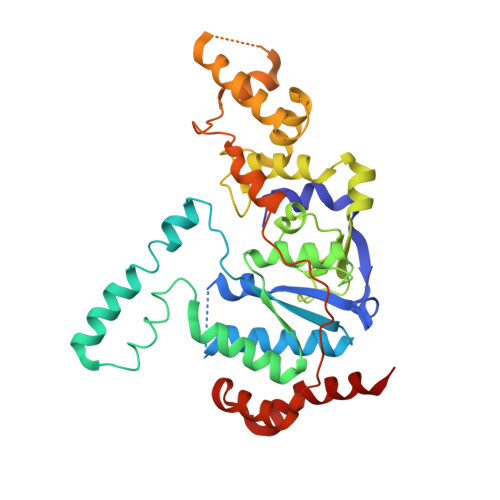
Human XPG nuclease structure, assembly, and activities with insights for neurodegeneration and cancer from pathogenic mutations.

Tsutakawa, S.E.Sarker, A.H.Ng, C.Arvai, A.S.Shin, D.S.Shih, B.Jiang, S.Thwin, A.C.Tsai, M.S.Willcox, A.Her, M.Z.Trego, K.S.Raetz, A.G.Rosenberg, D.Bacolla, A.Hammel, M.Griffith, J.D.Cooper, P.K.Tainer, J.A.

(2020) Proc Natl Acad Sci U S A 117: 14127-14138

  • DOI: https://doi.org/10.1073/pnas.1921311117
  • Primary Citation of Related Structures:  
    6VBH

  • PubMed Abstract: 

    Xeroderma pigmentosum group G (XPG) protein is both a functional partner in multiple DNA damage responses (DDR) and a pathway coordinator and structure-specific endonuclease in nucleotide excision repair (NER). Different mutations in the XPG gene ERCC5 lead to either of two distinct human diseases: Cancer-prone xeroderma pigmentosum (XP-G) or the fatal neurodevelopmental disorder Cockayne syndrome (XP-G/CS). To address the enigmatic structural mechanism for these differing disease phenotypes and for XPG's role in multiple DDRs, here we determined the crystal structure of human XPG catalytic domain (XPGcat), revealing XPG-specific features for its activities and regulation. Furthermore, XPG DNA binding elements conserved with FEN1 superfamily members enable insights on DNA interactions. Notably, all but one of the known pathogenic point mutations map to XPGcat, and both XP-G and XP-G/CS mutations destabilize XPG and reduce its cellular protein levels. Mapping the distinct mutation classes provides structure-based predictions for disease phenotypes: Residues mutated in XP-G are positioned to reduce local stability and NER activity, whereas residues mutated in XP-G/CS have implied long-range structural defects that would likely disrupt stability of the whole protein, and thus interfere with its functional interactions. Combined data from crystallography, biochemistry, small angle X-ray scattering, and electron microscopy unveil an XPG homodimer that binds, unstacks, and sculpts duplex DNA at internal unpaired regions (bubbles) into strongly bent structures, and suggest how XPG complexes may bind both NER bubble junctions and replication forks. Collective results support XPG scaffolding and DNA sculpting functions in multiple DDR processes to maintain genome stability.


  • Organizational Affiliation

    Molecular Biophysics and Integrated Bioimaging, Lawrence Berkeley National Laboratory, Berkeley, CA 94720; setsutakawa@lbl.gov jdg@med.unc.edu PKCooper@lbl.gov jatainer@gmail.com.


Macromolecules
Find similar proteins by:  (by identity cutoff)  |  3D Structure
Entity ID: 1
MoleculeChains Sequence LengthOrganismDetailsImage
DNA repair protein complementing XP-G cells,Flap endonuclease 1346Homo sapiensPyrococcus furiosusMutation(s): 0 
Gene Names: ERCC5ERCM2XPGXPGCfenfen-1PF1414
EC: 3.1
UniProt & NIH Common Fund Data Resources
Find proteins for O93634 (Pyrococcus furiosus (strain ATCC 43587 / DSM 3638 / JCM 8422 / Vc1))
Explore O93634 
Go to UniProtKB:  O93634
Find proteins for P28715 (Homo sapiens)
Explore P28715 
Go to UniProtKB:  P28715
PHAROS:  P28715
GTEx:  ENSG00000134899 
Entity Groups  
Sequence Clusters30% Identity50% Identity70% Identity90% Identity95% Identity100% Identity
UniProt GroupsP28715O93634
Sequence Annotations
Expand
  • Reference Sequence
Small Molecules
Ligands 1 Unique
IDChains Name / Formula / InChI Key2D Diagram3D Interactions
SO4
Query on SO4

Download Ideal Coordinates CCD File 
B [auth A]
C [auth A]
D [auth A]
E [auth A]
F [auth A]
B [auth A],
C [auth A],
D [auth A],
E [auth A],
F [auth A],
G [auth A],
H [auth A],
I [auth A],
J [auth A],
K [auth A],
L [auth A],
M [auth A],
N [auth A],
O [auth A],
P [auth A]
SULFATE ION
O4 S
QAOWNCQODCNURD-UHFFFAOYSA-L
Experimental Data & Validation

Experimental Data

  • Method: X-RAY DIFFRACTION
  • Resolution: 2.00 Å
  • R-Value Free: 0.244 
  • R-Value Work: 0.220 
  • R-Value Observed: 0.221 
  • Space Group: C 2 2 21
Unit Cell:
Length ( Å )Angle ( ˚ )
a = 64.438α = 90
b = 173.403β = 90
c = 101.608γ = 90
Software Package:
Software NamePurpose
HKL-2000data reduction
SCALEPACKdata scaling
PHASERphasing
PHENIXrefinement
PDB_EXTRACTdata extraction

Structure Validation

View Full Validation Report

Currently 6VBH does not have a validation slider image.


Entry History & Funding Information

Deposition Data


Funding OrganizationLocationGrant Number
National Institutes of Health/National Cancer Institute (NIH/NCI)United StatesP01CA92584
National Institutes of Health/National Institute of General Medical Sciences (NIH/NIGMS)United StatesR01 GM110387
National Institutes of Health/National Cancer Institute (NIH/NCI)United StatesR35 CA22043

Revision History  (Full details and data files)

  • Version 1.0: 2020-06-17
    Type: Initial release
  • Version 1.1: 2020-07-08
    Changes: Database references
  • Version 1.2: 2023-10-11
    Changes: Data collection, Database references, Refinement description