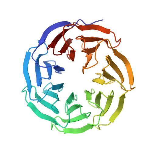
6UHZ

WDR5 in complex with Myc site fragment inhibitor


Experimental Data Snapshot

  • Method: X-RAY DIFFRACTION
  • Resolution: 1.26 Å
  • R-Value Free: 0.176 
  • R-Value Work: 0.152 
  • R-Value Observed: 0.153 

wwPDB Validation   3D Report Full Report


Ligand Structure Quality Assessment 


This is version 1.2 of the entry. See complete history


Literature

Discovery of WD Repeat-Containing Protein 5 (WDR5)-MYC Inhibitors Using Fragment-Based Methods and Structure-Based Design.

Chacon Simon, S.Wang, F.Thomas, L.R.Phan, J.Zhao, B.Olejniczak, E.T.Macdonald, J.D.Shaw, J.G.Schlund, C.Payne, W.Creighton, J.Stauffer, S.R.Waterson, A.G.Tansey, W.P.Fesik, S.W.

(2020) J Med Chem 63: 4315-4333

  • DOI: https://doi.org/10.1021/acs.jmedchem.0c00224
  • Primary Citation of Related Structures:  
    6UHY, 6UHZ, 6UIF, 6UIK, 6UJ4, 6UJH, 6UJJ, 6UJL, 6UOZ

  • PubMed Abstract: 

    The frequent deregulation of MYC and its elevated expression via multiple mechanisms drives cells to a tumorigenic state. Indeed, MYC is overexpressed in up to ∼50% of human cancers and is considered a highly validated anticancer target. Recently, we discovered that WD repeat-containing protein 5 (WDR5) binds to MYC and is a critical cofactor required for the recruitment of MYC to its target genes and reported the first small molecule inhibitors of the WDR5-MYC interaction using structure-based design. These compounds display high binding affinity, but have poor physicochemical properties and are hence not suitable for in vivo studies. Herein, we conducted an NMR-based fragment screening to identify additional chemical matter and, using a structure-based approach, we merged a fragment hit with the previously reported sulfonamide series. Compounds in this series can disrupt the WDR5-MYC interaction in cells, and as a consequence, we observed a reduction of MYC localization to chromatin.


  • Organizational Affiliation

    Department of Chemistry, Vanderbilt University, Nashville, Tennessee 37232, United States.


Macromolecules
Find similar proteins by:  (by identity cutoff)  |  3D Structure
Entity ID: 1
MoleculeChains Sequence LengthOrganismDetailsImage
WDR5
A, B
304Homo sapiensMutation(s): 0 
UniProt & NIH Common Fund Data Resources
Find proteins for P61964 (Homo sapiens)
Explore P61964 
Go to UniProtKB:  P61964
PHAROS:  P61964
GTEx:  ENSG00000196363 
Entity Groups  
Sequence Clusters30% Identity50% Identity70% Identity90% Identity95% Identity100% Identity
UniProt GroupP61964
Sequence Annotations
Expand
  • Reference Sequence
Small Molecules
Experimental Data & Validation

Experimental Data

  • Method: X-RAY DIFFRACTION
  • Resolution: 1.26 Å
  • R-Value Free: 0.176 
  • R-Value Work: 0.152 
  • R-Value Observed: 0.153 
  • Space Group: C 1 2 1
Unit Cell:
Length ( Å )Angle ( ˚ )
a = 134.374α = 90
b = 46.903β = 117.61
c = 112.462γ = 90
Software Package:
Software NamePurpose
HKL-2000data scaling
PHENIXrefinement
PDB_EXTRACTdata extraction
HKL-2000data reduction
PHASERphasing

Structure Validation

View Full Validation Report


Ligand Structure Quality Assessment 


Entry History & Funding Information

Deposition Data


Funding OrganizationLocationGrant Number
Department of Energy (DOE, United States)United StatesDE-AC02-06CH11357

Revision History  (Full details and data files)

  • Version 1.0: 2020-04-15
    Type: Initial release
  • Version 1.1: 2020-05-06
    Changes: Database references
  • Version 1.2: 2024-03-13
    Changes: Data collection, Database references