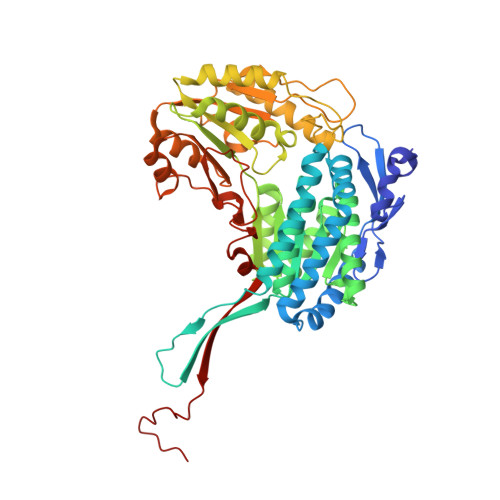
6U2X

Structure of ALDH7A1 mutant E399G complexed with NAD


Experimental Data Snapshot

  • Method: X-RAY DIFFRACTION
  • Resolution: 2.15 Å
  • R-Value Free: 0.233 
  • R-Value Work: 0.174 
  • R-Value Observed: 0.177 

wwPDB Validation   3D Report Full Report

Currently 6U2X does not have a validation slider image.


This is version 1.3 of the entry. See complete history


Literature

Structural analysis of pathogenic mutations targeting Glu427 of ALDH7A1, the hot spot residue of pyridoxine-dependent epilepsy.

Laciak, A.R.Korasick, D.A.Gates, K.S.Tanner, J.J.

(2020) J Inherit Metab Dis 43: 635-644

  • DOI: https://doi.org/10.1002/jimd.12184
  • Primary Citation of Related Structures:  
    6O4I, 6O4K, 6O4L, 6U2X

  • PubMed Abstract: 

    Certain loss-of-function mutations in the gene encoding the lysine catabolic enzyme aldehyde dehydrogenase 7A1 (ALDH7A1) cause pyridoxine-dependent epilepsy (PDE). Missense mutations of Glu427, especially Glu427Gln, account for ~30% of the mutated alleles in PDE patients, and thus Glu427 has been referred to as a mutation hot spot of PDE. Glu427 is invariant in the ALDH superfamily and forms ionic hydrogen bonds with the nicotinamide ribose of the NAD + cofactor. Here we report the first crystal structures of ALDH7A1 containing pathogenic mutations targeting Glu427. The mutant enzymes E427Q, Glu427Asp, and Glu427Gly were expressed in Escherichia coli and purified. The recombinant enzymes displayed negligible catalytic activity compared to the wild-type enzyme. The crystal structures of the mutant enzymes complexed with NAD + were determined to understand how the mutations impact NAD + binding. In the E427Q and E427G structures, the nicotinamide mononucleotide is highly flexible and lacks a defined binding pose. In E427D, the bound NAD + adopts a "retracted" conformation in which the nicotinamide ring is too far from the catalytic Cys residue for hydride transfer. Thus, the structures revealed a shared mechanism for loss of function: none of the variants are able to stabilise the nicotinamide of NAD + in the pose required for catalysis. We also show that these mutations reduce the amount of active tetrameric ALDH7A1 at the concentration of NAD + tested. Altogether, our results provide the three-dimensional molecular structural basis of the most common pathogenic variants of PDE and implicate strong (ionic) hydrogen bonds in the aetiology of a human disease.


  • Organizational Affiliation

    Department of Chemistry, University of Missouri, Columbia, Missouri.


Macromolecules
Find similar proteins by:  (by identity cutoff)  |  3D Structure
Entity ID: 1
MoleculeChains Sequence LengthOrganismDetailsImage
Alpha-aminoadipic semialdehyde dehydrogenase
A, B, C, D, E
A, B, C, D, E, F, G, H
513Homo sapiensMutation(s): 1 
Gene Names: ALDH7A1ATQ1
EC: 1.2.1.31 (PDB Primary Data), 1.2.1.3 (PDB Primary Data), 1.2.1.8 (PDB Primary Data)
UniProt & NIH Common Fund Data Resources
Find proteins for P49419 (Homo sapiens)
Explore P49419 
Go to UniProtKB:  P49419
PHAROS:  P49419
GTEx:  ENSG00000164904 
Entity Groups  
Sequence Clusters30% Identity50% Identity70% Identity90% Identity95% Identity100% Identity
UniProt GroupP49419
Sequence Annotations
Expand
  • Reference Sequence
Experimental Data & Validation

Experimental Data

  • Method: X-RAY DIFFRACTION
  • Resolution: 2.15 Å
  • R-Value Free: 0.233 
  • R-Value Work: 0.174 
  • R-Value Observed: 0.177 
  • Space Group: C 1 2 1
Unit Cell:
Length ( Å )Angle ( ˚ )
a = 155.27α = 90
b = 161.65β = 94.41
c = 158.8γ = 90
Software Package:
Software NamePurpose
Aimlessdata scaling
PHENIXrefinement
PDB_EXTRACTdata extraction
XDSdata reduction
PHASERphasing

Structure Validation

View Full Validation Report

Currently 6U2X does not have a validation slider image.


Entry History & Funding Information

Deposition Data


Funding OrganizationLocationGrant Number
National Institutes of Health/National Institute of General Medical Sciences (NIH/NIGMS)United StatesR01GM093123

Revision History  (Full details and data files)

  • Version 1.0: 2019-11-06
    Type: Initial release
  • Version 1.1: 2020-01-01
    Changes: Author supporting evidence
  • Version 1.2: 2020-05-20
    Changes: Database references
  • Version 1.3: 2023-10-11
    Changes: Data collection, Database references, Refinement description