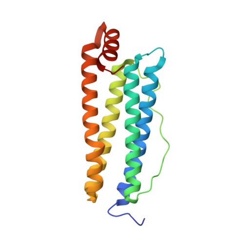
6TS1

Crystal structure of human L ferritin (HuLf) triple variant E60A-E61A-E64A Fe(III)-loaded for 60 minutes


Experimental Data Snapshot

  • Method: X-RAY DIFFRACTION
  • Resolution: 2.20 Å
  • R-Value Free: 0.238 
  • R-Value Work: 0.196 

wwPDB Validation   3D Report Full Report


This is version 1.2 of the entry. See complete history


Literature

Iron Biomineral Growth from the Initial Nucleation Seed in L-Ferritin.

Ciambellotti, S.Pozzi, C.Mangani, S.Turano, P.

(2020) Chemistry 26: 5770-5773

  • DOI: https://doi.org/10.1002/chem.202000064
  • Primary Citation of Related Structures:  
    6TR9, 6TRZ, 6TS0, 6TS1, 6TSA, 6TSF, 6TSJ, 6TSS, 6TSX

  • PubMed Abstract: 

    X-ray structures of homopolymeric human L-ferritin and horse spleen ferritin were solved by freezing protein crystals at different time intervals after exposure to a ferric salt and revealed the growth of an octa-nuclear iron cluster on the inner surface of the protein cage with a key role played by some glutamate residues. An atomic resolution view of how the cluster formation develops starting from a (μ 3 -oxo)tris[(μ 2 -glutamato-κO:κO')](glutamato-κO)(diaquo)triiron(III) seed is provided. The results support the idea that iron biomineralization in ferritin is a process initiating at the level of the protein surface, capable of contributing coordination bonds and electrostatic guidance.


  • Organizational Affiliation

    Magnetic Resonance Center (CERM), University of Florence, Sesto Fiorentino, 50019, Italy.


Macromolecules
Find similar proteins by:  (by identity cutoff)  |  3D Structure
Entity ID: 1
MoleculeChains Sequence LengthOrganismDetailsImage
Ferritin light chainA [auth AAA]175Homo sapiensMutation(s): 3 
Gene Names: FTL
UniProt & NIH Common Fund Data Resources
Find proteins for P02792 (Homo sapiens)
Explore P02792 
Go to UniProtKB:  P02792
PHAROS:  P02792
GTEx:  ENSG00000087086 
Entity Groups  
Sequence Clusters30% Identity50% Identity70% Identity90% Identity95% Identity100% Identity
UniProt GroupP02792
Sequence Annotations
Expand
  • Reference Sequence
Small Molecules
Ligands 4 Unique
IDChains Name / Formula / InChI Key2D Diagram3D Interactions
CD
Query on CD

Download Ideal Coordinates CCD File 
C [auth AAA]CADMIUM ION
Cd
WLZRMCYVCSSEQC-UHFFFAOYSA-N
GOL
Query on GOL

Download Ideal Coordinates CCD File 
D [auth AAA]GLYCEROL
C3 H8 O3
PEDCQBHIVMGVHV-UHFFFAOYSA-N
FE (Subject of Investigation/LOI)
Query on FE

Download Ideal Coordinates CCD File 
E [auth AAA]
F [auth AAA]
G [auth AAA]
H [auth AAA]
I [auth AAA]
E [auth AAA],
F [auth AAA],
G [auth AAA],
H [auth AAA],
I [auth AAA],
J [auth AAA],
K [auth AAA],
L [auth AAA],
M [auth AAA]
FE (III) ION
Fe
VTLYFUHAOXGGBS-UHFFFAOYSA-N
O
Query on O

Download Ideal Coordinates CCD File 
B [auth AAA]OXYGEN ATOM
O
XLYOFNOQVPJJNP-UHFFFAOYSA-N
Experimental Data & Validation

Experimental Data

  • Method: X-RAY DIFFRACTION
  • Resolution: 2.20 Å
  • R-Value Free: 0.238 
  • R-Value Work: 0.196 
  • Space Group: I 4 3 2
Unit Cell:
Length ( Å )Angle ( ˚ )
a = 151.222α = 90
b = 151.222β = 90
c = 151.222γ = 90
Software Package:
Software NamePurpose
REFMACrefinement
XDSdata reduction
SCALAdata scaling
MOLREPphasing

Structure Validation

View Full Validation Report


Entry History 

Deposition Data

Revision History  (Full details and data files)

  • Version 1.0: 2020-02-19
    Type: Initial release
  • Version 1.1: 2020-05-20
    Changes: Database references
  • Version 1.2: 2024-01-24
    Changes: Data collection, Database references, Derived calculations, Refinement description