6RT6

The YTH domain of YTHDC1 protein in complex with GGm6AC oligonucleotide


Experimental Data Snapshot

  • Method: X-RAY DIFFRACTION
  • Resolution: 1.46 Å
  • R-Value Free: 0.220 
  • R-Value Work: 0.206 
  • R-Value Observed: 0.207 

wwPDB Validation   3D Report Full Report


This is version 1.2 of the entry. See complete history


Literature

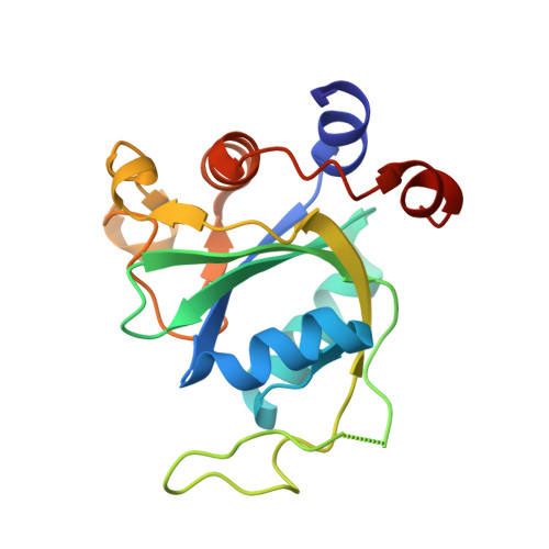
Flexible Binding of m6A Reader Protein YTHDC1 to Its Preferred RNA Motif.

Li, Y.Bedi, R.K.Wiedmer, L.Huang, D.Sledz, P.Caflisch, A.

(2019) J Chem Theory Comput 15: 7004-7014

  • DOI: https://doi.org/10.1021/acs.jctc.9b00987
  • Primary Citation of Related Structures:  
    6RT4, 6RT5, 6RT6, 6RT7

  • PubMed Abstract: 

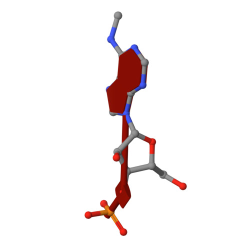
    N 6 -Methyladenosine (m 6 A) is the most prevalent chemical modification in human mRNAs. Its recognition by reader proteins enables many cellular functions, including splicing and translation of mRNAs. However, the binding mechanisms of m 6 A-containing RNAs to their readers are still elusive due to the unclear roles of m 6 A-flanking ribonucleotides. Here, we use a model system, YTHDC1 with its RNA motif 5'-G -2 G -1 (m 6 A)C +1 U +2 -3', to investigate the binding mechanisms by atomistic simulations, X-ray crystallography, and isothermal titration calorimetry. The experimental data and simulation results show that m 6 A is captured by an aromatic cage of YTHDC1 and the 3' terminus nucleotides are stabilized by cation-π-π interactions, while the 5' terminus remains flexible. Notably, simulations of unbound RNA motifs reveal that the methyl group of m 6 A and the 5' terminus shift the conformational preferences of the oligoribonucleotide to the bound-like conformation, thereby facilitating the association process. The binding mechanisms may help in the discovery of chemical probes against m 6 A reader proteins.


  • Organizational Affiliation

    Department of Biochemistry , University of Zurich , Winterthurerstrasse 190 , CH-8057 Zurich , Switzerland.


Macromolecules

Find similar proteins by:  (by identity cutoff)  |  3D Structure
Entity ID: 2
MoleculeChains Sequence LengthOrganismDetailsImage
YTH domain-containing protein 1
B, C
166Homo sapiensMutation(s): 0 
Gene Names: YTHDC1KIAA1966YT521
UniProt & NIH Common Fund Data Resources
Find proteins for Q96MU7 (Homo sapiens)
Explore Q96MU7 
Go to UniProtKB:  Q96MU7
PHAROS:  Q96MU7
GTEx:  ENSG00000083896 
Entity Groups  
Sequence Clusters30% Identity50% Identity70% Identity90% Identity95% Identity100% Identity
UniProt GroupQ96MU7
Sequence Annotations
Expand
  • Reference Sequence

Find similar nucleic acids by:  Sequence   |   3D Structure  

Entity ID: 1
MoleculeChains LengthOrganismImage
RNA (5'-R(*(6MZ)P*C)-3')A,
D [auth E]
4Homo sapiens
Sequence Annotations
Expand
  • Reference Sequence
Experimental Data & Validation

Experimental Data

  • Method: X-RAY DIFFRACTION
  • Resolution: 1.46 Å
  • R-Value Free: 0.220 
  • R-Value Work: 0.206 
  • R-Value Observed: 0.207 
  • Space Group: P 1 21 1
Unit Cell:
Length ( Å )Angle ( ˚ )
a = 39.913α = 90
b = 103.739β = 104.5
c = 41.983γ = 90
Software Package:
Software NamePurpose
PHENIXrefinement
XDSdata reduction
XSCALEdata scaling
PHASERphasing

Structure Validation

View Full Validation Report


Entry History 

Deposition Data

Revision History  (Full details and data files)

  • Version 1.0: 2019-11-27
    Type: Initial release
  • Version 1.1: 2019-12-18
    Changes: Database references
  • Version 1.2: 2024-01-24
    Changes: Data collection, Database references, Refinement description