6R63

Crystal structure of indoleamine 2,3-dioxygenase 1 (IDO1) in complex with ferric heme and MMG-0358


Experimental Data Snapshot

  • Method: X-RAY DIFFRACTION
  • Resolution: 2.89 Å
  • R-Value Free: 0.286 
  • R-Value Work: 0.234 
  • R-Value Observed: 0.238 

wwPDB Validation   3D Report Full Report


Ligand Structure Quality Assessment 


This is version 1.2 of the entry. See complete history


Literature

Inhibition Mechanisms of Indoleamine 2,3-Dioxygenase 1 (IDO1).

Rohrig, U.F.Reynaud, A.Majjigapu, S.R.Vogel, P.Pojer, F.Zoete, V.

(2019) J Med Chem 62: 8784-8795

  • DOI: https://doi.org/10.1021/acs.jmedchem.9b00942
  • Primary Citation of Related Structures:  
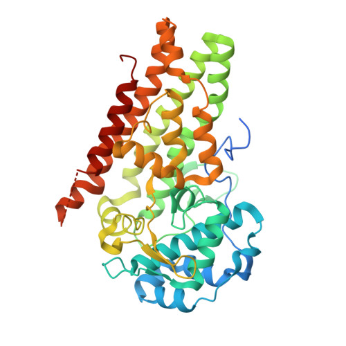
    6R63

  • PubMed Abstract: 

    Indoleamine 2,3-dioxygenase 1 (IDO1) catalyzes the rate-limiting step in the kynurenine pathway of tryptophan metabolism, which is involved in immunity, neuronal function, and aging. Its implication in pathologies such as cancer and neurodegenerative diseases has stimulated the development of IDO1 inhibitors. However, negative phase III clinical trial results of the IDO1 inhibitor epacadostat in cancer immunotherapy call for a better understanding of the role and the mechanisms of IDO1 inhibition. In this work, we investigate the molecular inhibition mechanisms of four known IDO1 inhibitors and of two quinones in detail, using different experimental and computational approaches. We also determine for the first time the X-ray structure of the highly efficient 1,2,3-triazole inhibitor MMG-0358. Based on our results and a comprehensive literature overview, we propose a classification scheme for IDO1 inhibitors according to their inhibition mechanism, which will be useful for further developments in the field.


  • Organizational Affiliation

    Molecular Modeling Group , SIB Swiss Institute of Bioinformatics , 1015 Lausanne , Switzerland.


Macromolecules
Find similar proteins by:  (by identity cutoff)  |  3D Structure
Entity ID: 1
MoleculeChains Sequence LengthOrganismDetailsImage
Indoleamine 2,3-dioxygenase 1
A, B
423Homo sapiensMutation(s): 0 
Gene Names: IDO1IDOINDO
EC: 1.13.11.52
UniProt & NIH Common Fund Data Resources
Find proteins for P14902 (Homo sapiens)
Explore P14902 
Go to UniProtKB:  P14902
PHAROS:  P14902
GTEx:  ENSG00000131203 
Entity Groups  
Sequence Clusters30% Identity50% Identity70% Identity90% Identity95% Identity100% Identity
UniProt GroupP14902
Sequence Annotations
Expand
  • Reference Sequence
Experimental Data & Validation

Experimental Data

  • Method: X-RAY DIFFRACTION
  • Resolution: 2.89 Å
  • R-Value Free: 0.286 
  • R-Value Work: 0.234 
  • R-Value Observed: 0.238 
  • Space Group: P 21 21 21
Unit Cell:
Length ( Å )Angle ( ˚ )
a = 90.91α = 90
b = 97.87β = 90
c = 119.235γ = 90
Software Package:
Software NamePurpose
PHENIXrefinement
XDSdata reduction
XDSdata scaling
PHASERphasing

Structure Validation

View Full Validation Report


Ligand Structure Quality Assessment 


Entry History 

Deposition Data

Revision History  (Full details and data files)

  • Version 1.0: 2019-10-02
    Type: Initial release
  • Version 1.1: 2019-10-23
    Changes: Data collection, Database references
  • Version 1.2: 2024-01-24
    Changes: Data collection, Database references, Derived calculations, Refinement description