6QIO

Ternary complex of FcRn ectodomain, FcRn binding optimised human serum albumin and the human growth hormone derivative somapacitan


Experimental Data Snapshot

  • Method: X-RAY DIFFRACTION
  • Resolution: 1.95 Å
  • R-Value Free: 0.226 
  • R-Value Work: 0.182 
  • R-Value Observed: 0.184 

wwPDB Validation   3D Report Full Report


Ligand Structure Quality Assessment 


This is version 2.1 of the entry. See complete history


Literature

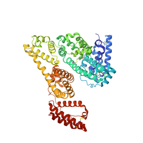
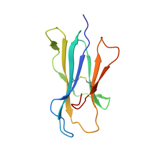
Identification of Binding Sites on Human Serum Albumin for Somapacitan, a Long-Acting Growth Hormone Derivative.

Johansson, E.Nielsen, A.D.Demuth, H.Wiberg, C.Schjodt, C.B.Huang, T.Chen, J.Jensen, S.Petersen, J.Thygesen, P.

(2020) Biochemistry 59: 1410-1419

  • DOI: https://doi.org/10.1021/acs.biochem.0c00019
  • Primary Citation of Related Structures:  
    6QIO, 6QIP

  • PubMed Abstract: 

    Somapacitan, a human growth hormone derivative that binds reversibly to albumin, was investigated for human serum albumin (HSA) and HSA domain binding. Isothermal titration calorimetry (ITC) binding profiles showed high-affinity binding (∼100-1000 nM) of one somapacitan molecule and low-affinity binding (∼1000-10000 nM) of one to two somapacitan molecules to HSA. The high-affinity site was identified in HSA domain III using size exclusion chromatography (SEC) and ITC. SEC studies showed that the neonatal Fc receptor shields one binding site for somapacitan, indicating its position in domain III. A crystal structure of somapacitan in complex with HSA optimized for neonatal Fc receptor binding, having four amino acid residue replacements, identified a low-affinity site in fatty acid-binding site 6 (domain II). Surface plasmon resonance (SPR) showed these replacements affect the kinetics of the high-affinity binding site. Furthermore, small-angle X-ray scattering and SPR brace two somapacitan-binding sites on HSA.


  • Organizational Affiliation

    Novo Nordisk A/S, Novo Nordisk Park 1, DK-2760 Måløv, Denmark.


Macromolecules
Find similar proteins by:  (by identity cutoff)  |  3D Structure
Entity ID: 1
MoleculeChains Sequence LengthOrganismDetailsImage
Serum albumin585Homo sapiensMutation(s): 4 
Gene Names: ALBGIG20GIG42PRO0903PRO1708PRO2044PRO2619PRO2675UNQ696/PRO1341
UniProt & NIH Common Fund Data Resources
Find proteins for P02768 (Homo sapiens)
Explore P02768 
Go to UniProtKB:  P02768
PHAROS:  P02768
GTEx:  ENSG00000163631 
Entity Groups  
Sequence Clusters30% Identity50% Identity70% Identity90% Identity95% Identity100% Identity
UniProt GroupP02768
Sequence Annotations
Expand
  • Reference Sequence
Find similar proteins by:  (by identity cutoff)  |  3D Structure
Entity ID: 2
MoleculeChains Sequence LengthOrganismDetailsImage
IgG receptor FcRn large subunit p51274Homo sapiensMutation(s): 0 
Gene Names: FCGRTFCRN
UniProt & NIH Common Fund Data Resources
Find proteins for P55899 (Homo sapiens)
Explore P55899 
Go to UniProtKB:  P55899
PHAROS:  P55899
GTEx:  ENSG00000104870 
Entity Groups  
Sequence Clusters30% Identity50% Identity70% Identity90% Identity95% Identity100% Identity
UniProt GroupP55899
Sequence Annotations
Expand
  • Reference Sequence
Find similar proteins by:  (by identity cutoff)  |  3D Structure
Entity ID: 3
MoleculeChains Sequence LengthOrganismDetailsImage
Beta-2-microglobulin105Homo sapiensMutation(s): 0 
Gene Names: B2MCDABP0092HDCMA22P
UniProt & NIH Common Fund Data Resources
Find proteins for P61769 (Homo sapiens)
Explore P61769 
Go to UniProtKB:  P61769
PHAROS:  P61769
GTEx:  ENSG00000166710 
Entity Groups  
Sequence Clusters30% Identity50% Identity70% Identity90% Identity95% Identity100% Identity
UniProt GroupP61769
Sequence Annotations
Expand
  • Reference Sequence
Experimental Data & Validation

Experimental Data

  • Method: X-RAY DIFFRACTION
  • Resolution: 1.95 Å
  • R-Value Free: 0.226 
  • R-Value Work: 0.182 
  • R-Value Observed: 0.184 
  • Space Group: P 21 21 21
Unit Cell:
Length ( Å )Angle ( ˚ )
a = 75.29α = 90
b = 108.12β = 90
c = 159.53γ = 90
Software Package:
Software NamePurpose
PHENIXrefinement
XDSdata reduction
XSCALEdata scaling
PHASERphasing

Structure Validation

View Full Validation Report


Ligand Structure Quality Assessment 


Entry History 

Deposition Data

Revision History  (Full details and data files)

  • Version 1.0: 2020-02-05
    Type: Initial release
  • Version 1.1: 2020-04-22
    Changes: Database references
  • Version 2.0: 2023-09-20
    Changes: Advisory, Atomic model, Data collection, Database references, Derived calculations, Polymer sequence, Refinement description, Source and taxonomy, Structure summary
  • Version 2.1: 2024-01-24
    Changes: Refinement description