6QC0

PCNA complex with Cdt2 C-terminal PIP-box peptide


Experimental Data Snapshot

  • Method: X-RAY DIFFRACTION
  • Resolution: 3.50 Å
  • R-Value Free: 0.252 
  • R-Value Work: 0.196 
  • R-Value Observed: 0.198 

wwPDB Validation   3D Report Full Report


This is version 1.0 of the entry. See complete history


Literature

Direct binding of Cdt2 to PCNA is important for targeting the CRL4Cdt2E3 ligase activity to Cdt1.

Hayashi, A.Giakoumakis, N.N.Heidebrecht, T.Ishii, T.Panagopoulos, A.Caillat, C.Takahara, M.Hibbert, R.G.Suenaga, N.Stadnik-Spiewak, M.Takahashi, T.Shiomi, Y.Taraviras, S.von Castelmur, E.Lygerou, Z.Perrakis, A.Nishitani, H.

(2018) Life Sci Alliance 1: e201800238-e201800238

  • DOI: https://doi.org/10.26508/lsa.201800238
  • Primary Citation of Related Structures:  
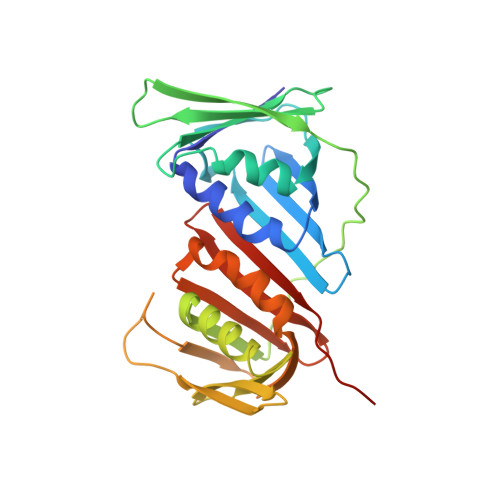
    6QC0, 6QCG

  • PubMed Abstract: 

    The CRL4 Cdt2 ubiquitin ligase complex is an essential regulator of cell-cycle progression and genome stability, ubiquitinating substrates such as p21, Set8, and Cdt1, via a display of substrate degrons on proliferating cell nuclear antigens (PCNAs). Here, we examine the hierarchy of the ligase and substrate recruitment kinetics onto PCNA at sites of DNA replication. We demonstrate that the C-terminal end of Cdt2 bears a PCNA interaction protein motif (PIP box, Cdt2 PIP ), which is necessary and sufficient for the binding of Cdt2 to PCNA. Cdt2 PIP binds PCNA directly with high affinity, two orders of magnitude tighter than the PIP box of Cdt1. X-ray crystallographic structures of PCNA bound to Cdt2 PIP and Cdt1 PIP show that the peptides occupy all three binding sites of the trimeric PCNA ring. Mutating Cdt2 PIP weakens the interaction with PCNA, rendering CRL4 Cdt2 less effective in Cdt1 ubiquitination and leading to defects in Cdt1 degradation. The molecular mechanism we present suggests a new paradigm for bringing substrates to the CRL4-type ligase, where the substrate receptor and substrates bind to a common multivalent docking platform to enable subsequent ubiquitination.


  • Organizational Affiliation

    Graduate School of Life Science, University of Hyogo, Kamigori, Japan.


Macromolecules
Find similar proteins by:  (by identity cutoff)  |  3D Structure
Entity ID: 1
MoleculeChains Sequence LengthOrganismDetailsImage
Proliferating cell nuclear antigenA,
B [auth C],
C [auth E]
263Homo sapiensMutation(s): 0 
Gene Names: PCNA
UniProt & NIH Common Fund Data Resources
Find proteins for P12004 (Homo sapiens)
Explore P12004 
Go to UniProtKB:  P12004
PHAROS:  P12004
GTEx:  ENSG00000132646 
Entity Groups  
Sequence Clusters30% Identity50% Identity70% Identity90% Identity95% Identity100% Identity
UniProt GroupP12004
Sequence Annotations
Expand
  • Reference Sequence

Find similar proteins by:  Sequence   |   3D Structure  

Entity ID: 2
MoleculeChains Sequence LengthOrganismDetailsImage
Denticleless protein homologD [auth B],
E [auth D],
F
14Homo sapiensMutation(s): 0 
UniProt & NIH Common Fund Data Resources
Find proteins for Q9NZJ0 (Homo sapiens)
Explore Q9NZJ0 
Go to UniProtKB:  Q9NZJ0
PHAROS:  Q9NZJ0
GTEx:  ENSG00000143476 
Entity Groups  
Sequence Clusters30% Identity50% Identity70% Identity90% Identity95% Identity100% Identity
UniProt GroupQ9NZJ0
Sequence Annotations
Expand
  • Reference Sequence
Experimental Data & Validation

Experimental Data

  • Method: X-RAY DIFFRACTION
  • Resolution: 3.50 Å
  • R-Value Free: 0.252 
  • R-Value Work: 0.196 
  • R-Value Observed: 0.198 
  • Space Group: P 61
Unit Cell:
Length ( Å )Angle ( ˚ )
a = 151.369α = 90
b = 151.369β = 90
c = 91.489γ = 120
Software Package:
Software NamePurpose
REFMACrefinement
XDSdata reduction
Aimlessdata scaling
MOLREPphasing

Structure Validation

View Full Validation Report


Entry History 

Deposition Data

Revision History  (Full details and data files)

  • Version 1.0: 2019-01-23
    Type: Initial release