6Q2S

Cryo-EM structure of RET/GFRa3/ARTN extracellular complex. The 3D refinement was applied with C2 symmetry.


Experimental Data Snapshot

  • Method: ELECTRON MICROSCOPY
  • Resolution: 3.80 Å
  • Aggregation State: PARTICLE 
  • Reconstruction Method: SINGLE PARTICLE 

wwPDB Validation   3D Report Full Report


This is version 1.1 of the entry. See complete history


Literature

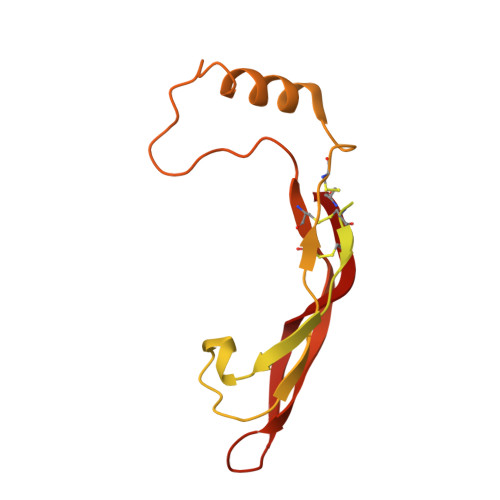
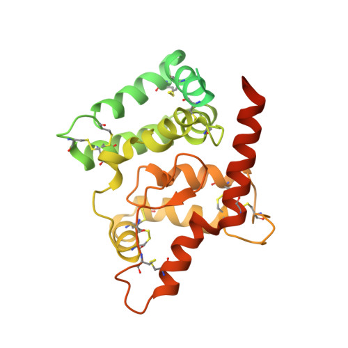
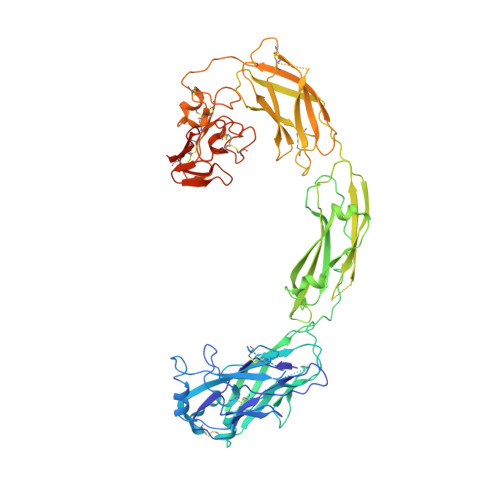
Cryo-EM analyses reveal the common mechanism and diversification in the activation of RET by different ligands.

Li, J.Shang, G.Chen, Y.J.Brautigam, C.A.Liou, J.Zhang, X.Bai, X.C.

(2019) Elife 8

  • DOI: https://doi.org/10.7554/eLife.47650
  • Primary Citation of Related Structures:  
    6Q2J, 6Q2N, 6Q2O, 6Q2R, 6Q2S

  • PubMed Abstract: 

    RET is a receptor tyrosine kinase (RTK) that plays essential roles in development and has been implicated in several human diseases. Different from most of RTKs, RET requires not only its cognate ligands but also co-receptors for activation, the mechanisms of which remain unclear due to lack of high-resolution structures of the ligand/co-receptor/receptor complexes. Here, we report cryo-EM structures of the extracellular region ternary complexes of GDF15/GFRAL/RET, GDNF/GFRα1/RET, NRTN/GFRα2/RET and ARTN/GFRα3/RET. These structures reveal that all the four ligand/co-receptor pairs, while using different atomic interactions, induce a specific dimerization mode of RET that is poised to bring the two kinase domains into close proximity for cross-phosphorylation. The NRTN/GFRα2/RET dimeric complex further pack into a tetrameric assembly, which is shown by our cell-based assays to regulate the endocytosis of RET. Our analyses therefore reveal both the common mechanism and diversification in the activation of RET by different ligands.


  • Organizational Affiliation

    Department of Biophysics, University of Texas Southwestern Medical Center, Dallas, United States.


Macromolecules
Find similar proteins by:  (by identity cutoff)  |  3D Structure
Entity ID: 1
MoleculeChains Sequence LengthOrganismDetailsImage
Ubiquitin-like protein SMT3,ArteminA,
D [auth B]
235Saccharomyces cerevisiaeHomo sapiens
This entity is chimeric
Mutation(s): 0 
Gene Names: SMT3ARTNEVN
UniProt & NIH Common Fund Data Resources
Find proteins for Q12306 (Saccharomyces cerevisiae (strain ATCC 204508 / S288c))
Explore Q12306 
Go to UniProtKB:  Q12306
Find proteins for Q5T4W7 (Homo sapiens)
Explore Q5T4W7 
Go to UniProtKB:  Q5T4W7
PHAROS:  Q5T4W7
GTEx:  ENSG00000117407 
Entity Groups  
Sequence Clusters30% Identity50% Identity70% Identity90% Identity95% Identity100% Identity
UniProt GroupsQ5T4W7Q12306
Sequence Annotations
Expand
  • Reference Sequence
Find similar proteins by:  (by identity cutoff)  |  3D Structure
Entity ID: 2
MoleculeChains Sequence LengthOrganismDetailsImage
GDNF family receptor alpha-3B [auth C],
E [auth D]
342Homo sapiensMutation(s): 0 
Gene Names: GFRA3UNQ339/PRO538/PRO3664
UniProt & NIH Common Fund Data Resources
Find proteins for O60609 (Homo sapiens)
Explore O60609 
Go to UniProtKB:  O60609
PHAROS:  O60609
GTEx:  ENSG00000146013 
Entity Groups  
Sequence Clusters30% Identity50% Identity70% Identity90% Identity95% Identity100% Identity
UniProt GroupO60609
Sequence Annotations
Expand
  • Reference Sequence
Find similar proteins by:  (by identity cutoff)  |  3D Structure
Entity ID: 3
MoleculeChains Sequence LengthOrganismDetailsImage
Proto-oncogene tyrosine-protein kinase receptor RetC [auth E],
F
617Homo sapiensMutation(s): 0 
Gene Names: RETCDHF12CDHR16PTCRET51
EC: 2.7.10.1
UniProt & NIH Common Fund Data Resources
Find proteins for P07949 (Homo sapiens)
Explore P07949 
Go to UniProtKB:  P07949
PHAROS:  P07949
GTEx:  ENSG00000165731 
Entity Groups  
Sequence Clusters30% Identity50% Identity70% Identity90% Identity95% Identity100% Identity
UniProt GroupP07949
Sequence Annotations
Expand
  • Reference Sequence
Small Molecules
Ligands 2 Unique
IDChains Name / Formula / InChI Key2D Diagram3D Interactions
NAG (Subject of Investigation/LOI)
Query on NAG

Download Ideal Coordinates CCD File 
G [auth C]
H [auth E]
I [auth E]
J [auth E]
K [auth E]
G [auth C],
H [auth E],
I [auth E],
J [auth E],
K [auth E],
L [auth E],
M [auth E],
R [auth D],
S [auth F],
T [auth F],
U [auth F],
V [auth F],
W [auth F],
X [auth F]
2-acetamido-2-deoxy-beta-D-glucopyranose
C8 H15 N O6
OVRNDRQMDRJTHS-FMDGEEDCSA-N
CA (Subject of Investigation/LOI)
Query on CA

Download Ideal Coordinates CCD File 
AA [auth F]
BA [auth F]
N [auth E]
O [auth E]
P [auth E]
AA [auth F],
BA [auth F],
N [auth E],
O [auth E],
P [auth E],
Q [auth E],
Y [auth F],
Z [auth F]
CALCIUM ION
Ca
BHPQYMZQTOCNFJ-UHFFFAOYSA-N
Experimental Data & Validation

Experimental Data

  • Method: ELECTRON MICROSCOPY
  • Resolution: 3.80 Å
  • Aggregation State: PARTICLE 
  • Reconstruction Method: SINGLE PARTICLE 
EM Software:
TaskSoftware PackageVersion
MODEL REFINEMENTPHENIX
RECONSTRUCTIONRELION

Structure Validation

View Full Validation Report


Entry History 

Deposition Data

Revision History  (Full details and data files)

  • Version 1.0: 2019-10-02
    Type: Initial release
  • Version 1.1: 2020-07-29
    Type: Remediation
    Reason: Carbohydrate remediation
    Changes: Data collection, Derived calculations, Structure summary